The Delta Dispatch
Ejection Trauma: What First Responders Need to Know About High-Impact MVCs
Ejection trauma is one of the most catastrophic outcomes of a motor vehicle collision. For first responders, understanding how to assess, stabilize, and prioritize care for these high-impact patients is critical. Here’s how to stay calm, organized, and effective in the moments that matter most.
When a person is ejected from a vehicle during a crash, the outcome is almost always severe. Ejection trauma represents one of the most catastrophic mechanisms of injury emergency responders will encounter. Whether partial or complete, being thrown from a vehicle amplifies the forces acting on the body—turning an already violent impact into a multi-system trauma event.
What Is Ejection Trauma?
Ejection trauma occurs when a vehicle occupant is completely or partially thrown out of the vehicle during a collision. This typically happens when seat belts are not worn or fail, or when the force of impact breaches the vehicle’s structure.
There are two main types:
Partial ejection – The patient is partially expelled, often trapped by the seat belt or vehicle frame, resulting in crushing or shearing injuries.
Complete ejection – The patient is fully thrown clear of the vehicle, often resulting in secondary impacts with the ground or other objects.
Mechanism of Injury (MOI)
Ejection trauma is classified as a high-mechanism MOI, and responders should immediately suspect:
Severe blunt trauma
Multiple fractures
Spinal, head, and internal injuries
Airway compromise or shock
The energy transfer doesn’t stop at the first impact—the patient often experiences multiple points of contact:
The initial crash inside the vehicle
Contact with the vehicle structure on exit
Impact with the ground or another surface
Potential secondary impact with objects (trees, poles, other vehicles)
Each of these contributes to a complex and unpredictable injury pattern.
Common Injuries Associated with Ejection
Responders should anticipate multi-system trauma, including:
Head and neck injuries: Concussions, skull fractures, traumatic brain injuries (TBI)
Spinal trauma: High risk of spinal fractures or cord damage due to rotational forces
Thoracic injuries: Flail chest, pneumothorax, hemothorax, cardiac contusions
Abdominal trauma: Liver or spleen lacerations, internal bleeding
Orthopedic injuries: Long bone fractures, pelvic fractures, amputations
Soft tissue injuries: Severe abrasions, degloving injuries, lacerations
Scene Assessment
When responding to an MVC with possible ejection, scene safety and situational awareness come first.
Scan the entire area—patients may be thrown significant distances or hidden in brush, ditches, or behind obstacles.
Note patient position relative to the vehicle; it offers key clues about trajectory and energy transfer.
Look for additional hazards: leaking fuel, oncoming traffic, downed power lines.
Patient Management in the Field
Ejected patients require rapid, systematic assessment and early activation of advanced care.
1. Scene Safety & Triage
Request additional units and air transport early.
Perform a global scan for multiple patients.
2. Primary Survey (Check, Call, Care)
Airway: Suspect spinal injury—manually stabilize head and neck, use jaw-thrust if needed.
Breathing: Expose the chest, check for symmetry and adequate air movement.
Circulation: Control major bleeding immediately. Prioritize rapid identification of life-threatening hemorrhage.
3. Secondary Survey
Head-to-toe assessment, noting deformities, contusions, abrasions, punctures, burns, tenderness, lacerations, and swelling (DCAP-BTLS).
Monitor vitals frequently—changes can indicate internal bleeding or decompensating shock.
Maintain spinal motion restriction (SMR) if indicated.
4. Shock Management
Treat for hypovolemia: position appropriately, keep warm, and prepare for rapid transport.
Oxygen administration and airway management should be prioritized.
The Role of Prehospital Responders
As a first responder, your calm and methodical approach can make all the difference. Recognizing the severity of ejection trauma early allows for:
Prioritizing rapid extrication and transport
Effective communication with dispatch and incoming EMS
Early notification of trauma centers
Ejection = Severe Trauma Until Proven Otherwise.
Never underestimate the forces involved—patients may appear stable initially but can deteriorate quickly.
Key Takeaways
Ejection trauma is one of the deadliest outcomes of an MVC.
Always assume multi-system trauma and spinal involvement.
Focus on airway, breathing, and circulation while minimizing movement.
Rapid transport and early hospital notification are essential.
Scene safety, teamwork, and clear communication are critical under pressure.
Final Thoughts
Every ejection call reminds responders why scene assessment and mechanism recognition matter so much. Behind every statistic is a human life—one that depends on the calm, structured, and skilled actions of those first on scene. Whether you’re an Advanced First Aider, EMR, or paramedic student, understanding ejection trauma prepares you to manage chaos with confidence and compassion.
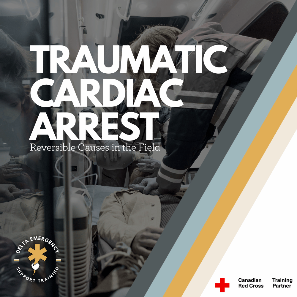
Traumatic Cardiac Arrest: What Every Professional Responder Should Know
Traumatic cardiac arrest is one of the most demanding situations a responder can face. This in-depth guide explains how to rapidly identify reversible causes, manage airway and bleeding, and provide effective field care to improve survival chances in severe trauma cases.
Traumatic cardiac arrest (TCA) represents one of the most challenging situations in prehospital care. Outcomes are often poor, but when managed rapidly and effectively, survival with good neurological recovery is possible. This blog explores what TCA is, its causes, assessment and treatment priorities, and best practices for professional responders in Canada.
What Is Traumatic Cardiac Arrest?
A traumatic cardiac arrest occurs when a patient’s heart stops as a direct result of major trauma. This trauma can be blunt (such as falls or motor vehicle collisions) or penetrating (such as gunshots or stab wounds).
Unlike medical cardiac arrests, TCA often stems from reversible physical causes rather than primary heart dysfunction. The most common reversible causes include:
Severe hemorrhage (uncontrolled bleeding)
Hypovolemia (loss of circulating volume)
Hypoxia (insufficient oxygen supply)
Tension pneumothorax (collapsed lung causing pressure on the heart)
Cardiac tamponade (blood filling the pericardial sac and compressing the heart)
How Common Is TCA and What Are the Survival Trends?
Traumatic cardiac arrests are less common than medical arrests but are often more complex. Historically, survival rates have been extremely low—typically below 2%. However, in cases where the underlying cause is rapidly identified and corrected, patients can and do survive, especially when there are early signs of life or immediate advanced care.
Outcomes tend to be more favorable in penetrating trauma compared to blunt trauma, largely due to the speed at which reversible causes can be treated.
Key Priorities in Managing TCA
When seconds matter, responders must focus on interventions that directly address reversible causes. The following priorities guide professional care:
PriorityActionPurposeControl Hemorrhage / HypovolemiaApply direct pressure, tourniquets, wound packing, or pelvic binders.Prevent further blood loss and restore perfusion.Airway and OxygenationClear the airway and ventilate effectively. Use supraglottic or advanced airways if trained and authorized.Correct hypoxia, one of the leading causes of TCA.Relieve Mechanical ObstructionsPerform needle decompression for tension pneumothorax or use a chest seal for open chest wounds.Restore normal breathing and heart function.High-Quality CPROnly once reversible causes are being managed. Focus on effective compressions with minimal interruptions.Maintain perfusion if reversible causes have been addressed.Rapid TransportTransport quickly to a trauma centre if signs of life are present.Definitive care is essential for survival.
Trauma Type and Approach
Penetrating Trauma
Penetrating injuries, such as gunshots or stabbings, can cause localized damage but often have a shorter timeline to intervention. Quick chest decompression, bleeding control, and airway management can make a significant difference in survival outcomes.
Blunt Trauma
Blunt trauma, such as from high-impact collisions or falls, often causes widespread internal injuries and massive bleeding. Survival rates are generally lower, and early recognition of futility is crucial when there are no signs of life and prolonged downtime.
Canadian Best Practices and Field Considerations
Canadian EMS and paramedic protocols emphasize a systematic, cause-driven approach to TCA management:
Address all reversible causes simultaneously.
Control bleeding, relieve pneumothorax, and manage airway without delay.Obtain IV/IO access and administer fluids as indicated for hypovolemia.
Perform bilateral needle decompressions if chest trauma is suspected.
Apply pelvic binders when pelvic fractures or major lower-body trauma are likely.
Minimize scene time. Rapid transport to a trauma centre remains a priority.
Epinephrine is often deprioritized, as it offers limited benefit when the heart has stopped due to physical injury rather than electrical failure.
Termination of Resuscitation (TOR) may be considered if there are no signs of life, asystole, and extended downtime with no reversible cause found.
Common Challenges for Responders
Traumatic cardiac arrests demand rapid thinking and precise teamwork. Common challenges include:
Identifying reversible vs. irreversible causes. It can be difficult to determine whether the arrest is trauma-induced or secondary to a medical event.
Time pressure. Every second counts, especially when massive hemorrhage or tension pneumothorax is suspected.
Limited prehospital tools. Not all regions permit advanced procedures like thoracotomy or pericardiocentesis.
Balancing interventions. CPR may worsen bleeding if hemorrhage is uncontrolled, so priorities must be reassessed continually.
Transport logistics. Long transport times may influence whether resuscitation efforts continue.
Step-By-Step Approach to Traumatic Cardiac Arrest
Ensure scene safety and don PPE.
Rapidly assess mechanism and timeline (blunt vs. penetrating, time of arrest, signs of life).
Control massive bleeding immediately. Use direct pressure, tourniquets, or wound packing.
Manage the airway and ventilate. Provide oxygen and ensure adequate chest rise.
Relieve chest pressure. Perform needle decompression and apply chest seals as indicated.
Provide fluid resuscitation. Administer IV or IO fluids per protocol.
Perform CPR if appropriate. Begin once reversible causes are actively addressed.
Transport rapidly to a trauma facility if ROSC occurs or if the patient shows any signs of life.
Consider termination if no response and all reversible causes have been managed.
Why TCA Differs from Medical Cardiac Arrest
In medical cardiac arrest, the primary issue is usually electrical or metabolic failure of the heart. In TCA, the heart often stops because of physical factors such as massive bleeding or chest injury. As a result, standard interventions like defibrillation or epinephrine are secondary to correcting the underlying mechanical or circulatory issues.
In short — treat the cause, not just the rhythm.
Realistic Scenarios
Penetrating Chest Trauma: A patient with a gunshot wound to the chest is pulseless on arrival. Responders immediately apply a chest seal, decompress the chest, manage the airway, and control external bleeding.
Blunt Pelvic Trauma: Following a vehicle collision, the patient has severe pelvic instability and no pulse. A pelvic binder is applied, external bleeding controlled, airway secured, and fluids initiated during transport.
Final Thoughts
Traumatic cardiac arrest demands composure, coordination, and deep understanding of trauma physiology. While survival rates remain low, outcomes improve significantly when responders rapidly identify and correct reversible causes.
Every second counts — and with the right interventions, professional responders can give trauma patients their best chance at survival.
Treating Gunshot Wounds: A Professional Responder’s Guide
Gunshot wounds are life-threatening emergencies that require quick, precise action. This guide for professional responders covers wound assessment, bleeding control, chest seal application for thoracic injuries, and key steps for safe transport to trauma centers.
Gunshot wounds (GSWs) are among the most critical emergencies a responder can encounter. Severity depends on bullet type, trajectory, and location, making rapid assessment and precise intervention essential. Professional responders must be prepared to manage everything from external bleeding to life-threatening internal injuries.
Gunshot Wounds in Canada: Context & Stats
Understanding the prevalence and patterns of firearm injuries in Canada provides important context:
Between 2016–2020, Canada saw over 4,000 deaths and thousands of hospitalizations due to firearm injuries.
Fatal injuries are most often suicides (~72%), followed by homicides (~23%), with unintentional injuries and law enforcement incidents making up smaller percentages.
Non-fatal injuries frequently result from assault or accidental shootings, highlighting the need for skilled first responders.
Young adult males (20–34) are most commonly affected, emphasizing the importance of rapid intervention in high-risk scenarios.
Scene Safety & Initial Approach
Before approaching the patient:
Ensure scene safety. Gunshot incidents may occur in violent settings—confirm the area is secure and involve law enforcement if necessary.
Use proper PPE. Gloves, eye protection, and body protection may be required depending on the scene.
Triage and rapid assessment. Identify life-threatening injuries first, especially massive bleeding or airway compromise.
Primary Survey: C-ABCDE Approach
Professional responders often use a C-ABCDE approach, prioritizing massive bleeding first:
C – Massive Hemorrhage: Apply direct pressure, hemostatic dressings, or tourniquets (for limbs). Junctional areas may require packing or specialized devices.
A – Airway: Ensure airway is clear; use adjuncts if necessary. Be alert for obstructions from blood, tissue, or debris.
B – Breathing: Evaluate for chest injuries. Gunshot wounds to the thorax can cause pneumothorax, tension pneumothorax, or hemothorax.
C – Circulation: Monitor vitals, assess for shock, establish IV access if in scope, and be ready to administer fluids per protocol.
D – Disability: Evaluate neurological status (AVPU or GCS) and look for signs of head or spinal injury.
E – Exposure & Environment: Fully assess injuries while keeping the patient warm and preserving dignity.
Treating Gunshot Wounds by Location
Extremities (Arms & Legs)
Risks: Severe bleeding, fractures, nerve damage.
Intervention: Direct pressure, tourniquets for limbs, immobilize fractures, monitor for shock.
Junctional Areas (Groin, Axilla, Neck)
Risks: Major vessel injury; uncontrolled bleeding.
Intervention: Use pressure and hemostatic dressings; junctional tourniquet if available.
Chest
Chest injuries are especially dangerous due to risk of lung collapse, cardiac injury, or massive internal bleeding.
Signs: Difficulty breathing, sucking chest wound, rapid deterioration, shock.
Intervention:
Chest Seal: Apply to open chest wounds to prevent air entry and reduce risk of tension pneumothorax. Venting seals allow trapped air to escape.
Oxygen Support: Provide high-flow oxygen if trained.
Positioning: Keep the patient upright if comfortable to assist breathing.
Rapid Transport: Patients with chest GSWs require urgent trauma care.
Abdomen & Pelvis
Risks: Internal organ damage, significant bleeding.
Intervention: Cover exposed organs with sterile, moist dressings, avoid manipulation, monitor vitals, rapid transport.
Head & Neck
Risks: Airway compromise, brain injury, severe bleeding.
Intervention: Protect cervical spine, manage airway, control bleeding, rapid transport.
Special Considerations
Bullet Energy & Cavitation: High-velocity bullets create more tissue damage due to cavitation.
Shock & Hypovolemia: Internal bleeding may be hidden—monitor vitals closely and maintain patient warmth.
Infection Prevention: Stabilize the patient, then follow protocols for wound cleaning, antibiotics, and tetanus prophylaxis.
Documentation & Legal Obligations: In Canada, gunshot injuries often must be reported to authorities. Preserve evidence while delivering care.
Tools & Equipment for Responders
Hemostatic dressings / pressure dressings
Tourniquets for limbs
Chest seals for sucking chest wounds
Airway adjuncts (OPA/NPA)
IV fluids and monitoring equipment (per protocol)
High-flow oxygen
PPE for responder safety
Transport & Handover
Maintain bleeding control, airway, and vital signs during transport.
Provide a clear handover to receiving trauma center: wound location, number of shots, interventions performed, and patient vitals.
Summary
Gunshot wounds are complex and life-threatening. Professional responders must:
Rapidly control bleeding and manage airways.
Use chest seals for thoracic injuries.
Monitor for shock and hidden injuries.
Provide rapid transport to trauma centers with thorough documentation.
Proper training, situational awareness, and decisive action can significantly improve survival and reduce complications from GSWs.
Managing Intoxicated Patients in the Field: An EMR Guide
As an Emergency Medical Responder (EMR), you’re trained to provide care in unpredictable situations. One of the more challenging scenarios is responding to patients under the influence of alcohol or drugs. Intoxicated patients can present with unpredictable behavior, altered physiology, and a higher risk of injury—making careful assessment and management critical.
At Delta Emergency Support Training, we emphasize both safety and effective patient care. Here’s a detailed guide for EMRs on safely responding to intoxicated patients, aligned with Alberta Health Services (AHS) guidelines.
Understanding Intoxication
Intoxication can result from alcohol, prescription medications, or illicit drugs. Common effects include:
Altered level of consciousness
Slurred speech or impaired coordination
Agitation, aggression, or unpredictable behavior
Nausea, vomiting, or respiratory depression
Increased risk of trauma from falls, fights, or accidents
Masked pain: Alcohol can dull pain perception, making trauma patients unreliable reporters of injury severity
Mimics: Hyperglycemia or hypoglycemia can appear similar to intoxication, making blood glucose assessment crucial
Recognizing the signs early helps EMRs anticipate complications and prioritize patient and responder safety.
Safety First: Protecting Yourself and Others
Intoxicated patients can be unpredictable. Before providing care, ensure:
Scene safety: Survey for weapons, hazards, or bystanders that may interfere.
Backup: If the patient is aggressive, call law enforcement or additional responders.
Personal protective equipment (PPE): Gloves, eye protection, and masks may be necessary.
Distance and positioning: Maintain a safe stance and keep an exit route clear.
Never compromise your safety—intoxicated patients may act impulsively, and an aggressive patient can escalate quickly.
Patient Assessment
Assessment of an intoxicated patient may be complicated by their altered state. EMRs should focus on:
Responsiveness: Determine if the patient is fully conscious, verbally responsive, physically responsive, or unresponsive.
Airway, Breathing, Circulation (ABCs): Alcohol and drugs can depress breathing and circulation.
Be prepared to clear the airway using suction if the patient vomits.
Airway adjuncts (oropharyngeal or nasopharyngeal airway) may be required if the patient’s airway is compromised but spontaneous breathing is present.
Vital signs: Monitor heart rate, blood pressure, respiratory rate, oxygen saturation, and blood glucose.
Hyperglycemia or hypoglycemia can mimic intoxication and significantly affect patient management.
Secondary survey: Look for trauma, especially since alcohol can mask pain. A patient may not report broken bones, internal injuries, or head trauma. Monitor carefully for subtle signs.
Common Challenges and How to Manage Them
Aggression or agitation: Stay calm, use a non-confrontational tone, and avoid physical confrontation unless absolutely necessary.
Vomiting or aspiration risk: Use suction immediately if needed, and position the patient in the recovery positionif unconscious but breathing.
Altered decision-making: Intoxicated patients may refuse care. Document refusals and use implied consent in life-threatening situations.
Polysubstance use: Patients may have multiple substances in their system, complicating presentation. Monitor closely for unexpected symptoms.
Trauma considerations: Be extra vigilant for head injuries, internal bleeding, or fractures. Pain may be unreliable, so rely on vital signs, visual assessment, and mechanism of injury.
Glucose considerations: Rapidly check blood glucose in any altered patient; treating hypoglycemia or hyperglycemia may resolve the presentation entirely.
Communication Tips
Use simple, clear language.
Speak slowly and calmly to reduce confusion.
Avoid judgmental language—patients respond better when treated respectfully.
Keep bystanders informed, but focus on patient care.
Transport Considerations
Monitor continuously: Intoxicated patients can deteriorate rapidly.
Secure safely: If aggressive, use restraints only per local protocols and monitor the patient closely.
Communicate with receiving facility: Provide details on level of intoxication, suspected substances, vital signs, airway interventions (suction or adjuncts used), trauma concerns, and any interventions performed.
Red Flags: When to Call for Immediate Backup
Unresponsiveness or decreased level of consciousness
Difficulty breathing or abnormal airway sounds
Vomiting with risk of aspiration
Seizures or convulsions
Severe trauma, especially head or internal injuries
Aggressive or violent behavior that cannot be safely managed
Hypoglycemia or hyperglycemia causing altered mental status
Final Thoughts
Responding to intoxicated patients requires a balance of emergency care skills, safety awareness, and empathy. Alcohol or drug use can mask pain and complicate assessment, and abnormal blood glucose levels can mimic intoxication. With proper airway management, suction, airway adjuncts, and glucose monitoring, EMRs can provide effective care safely.
At Delta Emergency Support Training, our courses prepare responders to handle complex patient scenarios, including intoxicated and trauma patients so you can provide care with confidence, skill, and safety.
Distracting Injuries: Pulling Focus and Masking Pain
In trauma care, some injuries grab all the attention—but they aren’t always the deadliest. Distracting injuries can mislead responders and mask life-threatening conditions. Here’s what every first responder needs to know about spotting them, staying systematic, and keeping patient safety the top priority.
When trauma calls come in, it’s easy to fixate on the most obvious wound, the open fracture, the deep laceration, the heavy bleeding. But those are often distracting injuries: visible traumas that can obscure other, more dangerous threats. Recognizing them and continuing to assess systematically is what separates a good response from a lifesaving one.
What Is a Distracting Injury?
A distracting injury is one that draws your attention so strongly that both responders and patients may overlook other serious but less obvious issues. In paramedic protocols, these injuries can make a patient “unreliable” for clinical clearance of the cervical spine because they may mask neck pain or tenderness. As a result, spinal precautions should be maintained unless more definitive assessment rules them out.
Why They Matter
The danger of distracting injuries is twofold:
They pull focus – A gruesome fracture or large bleed grabs everyone’s attention, often overshadowing a more subtle but life-threatening issue like a compromised airway or spinal injury.
They mask pain – A patient in severe pain from a major injury may not report or even notice neck or back pain, leading responders to miss critical red flags for spinal trauma.
Evidence-Based Insights
Studies show distracting injuries are often upper-torso or limb injuries that change how a patient perceives or reports pain:
Heffernan et al. (2005) found that some patients with cervical spine fractures had no neck tenderness—but all had upper torso injuries like chest trauma, which likely distracted from the pain.
Common Examples
Long bone fractures (femur, humerus)
Major burns
Crushing or degloving wounds
Profuse bleeding or amputations
Severe chest or upper torso injuries
First Responder Priorities
Stick to the primary survey (ABCs)
No matter how distracting a wound looks, airway, breathing, and circulation must always come first.Protect the spine when in doubt
If a distracting injury is present, assume the patient cannot reliably clear their spine. Apply spinal precautions unless imaging or protocols say otherwise.Reassess constantly
A distracting injury may not only occupy the patient’s attention but yours as well. Don’t let tunnel vision take over.Think systematically, not emotionally
The most dramatic injuries are not always the deadliest. Quiet but hidden injuries like internal bleeding or spinal cord compromise, may be far more dangerous.
The Bottom Line
Distracting injuries are more than just dramatic wounds—they can hide life-threatening problems and mislead both responders and patients. The best protection is discipline: follow a structured assessment, protect the spine when necessary, and never let your focus shift entirely to the injury that looks worst. A calm, systematic approach can make the difference between managing trauma and saving a life.
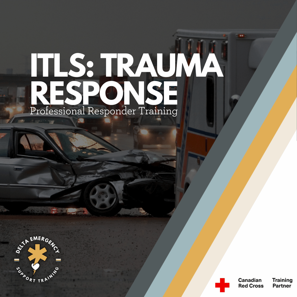
International Trauma Life Support: A Structural Approach to Trauma
Need to take ITLS but not sure what it entails? We’ll break it down for you. International Trauma Life Support (ITLS) gives professional responders the tools to quickly assess, stabilize, and manage trauma patients in high-stress situations. From airway management to bleeding control and spinal care, this course equips you with practical, hands-on skills that could save lives when every second counts.
When emergencies happen, paramedics, firefighters, and advanced first aid responders, having the right tools and knowledge can make the difference between life and death. International Trauma Life Support (ITLS) is a specialized training program that equips professional responders with the skills to rapidly assess, stabilize, and manage trauma patients in high-pressure situations.
If you’re a responder required to take ITLS, here’s what you need to know.
The Purpose of ITLS
ITLS focuses on a structured approach to trauma care. The goal is to give responders a proven framework for:
Quickly identifying life-threatening injuries.
Performing rapid, effective interventions.
Preventing further harm while preparing for transport.
Working within professional guidelines and medical protocols.
This training bridges the gap between classroom learning and the unpredictable realities of trauma scenes.
Core Learning in ITLS
Here’s a breakdown of the key skill areas covered in ITLS and what they mean in practice.
1. Patient Assessment & Initial Survey
Responders learn a systematic way to approach every trauma patient:
Hazards, Environment, MOI, Patient count and Assistance.
Primary survey (C-Spine, LOC, ABCs, RBS,).
Identifying life threats in the first few minutes.
Why it matters: A structured assessment ensures nothing is missed under pressure.
2. Airway Management & Adjuncts
Maintaining an open airway is priority number one. ITLS covers:
Manual maneuvers (jaw thrust, head-tilt chin-lift).
Airway adjuncts (OPA, NPA).
Advanced options (supraglottic airways, endotracheal intubation where permitted).
Recognizing and managing obstructions.
Why it matters: Without a clear airway, nothing else matters — oxygen can’t reach the body.
3. Breathing & Ventilation Support
Responders are trained to:
Recognize respiratory distress and failure.
Provide rescue breaths and ventilations with a bag-valve mask.
Use oxygen.
Identify and treat life-threatening chest injuries (tension pneumothorax, sucking chest wound).
Why it matters: Oxygen delivery is essential to survival, recognizing and treating chest trauma is lifesaving.
4. Circulation & Hemorrhage Control
Stopping major bleeding and supporting circulation includes:
Direct pressure and wound packing.
Tourniquet application.
Recognizing shock and initiating care.
Intravenous (IV) or intraosseous (IO) fluid administration (where scope allows).
Why it matters: Uncontrolled bleeding is one of the top causes of preventable trauma deaths.
5. Spinal Motion Restriction (SMR)
Trauma often comes with potential spinal injuries. ITLS emphasizes:
Patient assessment for spinal risk.
Manual stabilization and cervical collars.
Log rolls and backboard use.
Updated evidence-based approaches to SMR.
Why it matters: Proper handling prevents permanent paralysis from secondary injury.
6. Head & Chest Trauma Management
Specific scenarios covered include:
Managing traumatic brain injuries and increased intracranial pressure.
Recognizing chest trauma (flail chest, open chest wounds).
Interventions like occlusive dressings, fracture stabilization and deadly bleed control.
Why it matters: These injuries are time-sensitive and require immediate, precise intervention.
7. Protocols & Teamwork
Beyond individual skills, ITLS teaches responders to:
Work under medical protocols and established guidelines.
Communicate effectively in teams.
Prioritize transport decisions (“load and go” vs. “stay and play”).
Why it matters: Trauma care is never solo — effective teamwork saves lives.
Why Professional Responders Need ITLS
For those moving into advanced roles (Advanced First Aid, EMR, PCP, paramedic), ITLS is often a required certification. It ensures all responders use the same language, protocols, and structured assessment, which makes coordination seamless during emergencies.
With ITLS, you’re not just checking a box — you’re developing:
Confidence in high-stress scenarios.
Competence in trauma management skills.
Credibility as a trained professional who can handle life-threatening situations.
Final Takeaway
If you’re required to take ITLS, think of it as more than a course — it’s a survival toolkit for the real world of trauma response. You’ll learn how to rapidly assess patients, manage airways, stop life-threatening bleeds, stabilize spines, and follow trauma protocols effectively.
For professional responders, ITLS is not just training. It’s a commitment to ensuring that when seconds count, you can act decisively, safely, and in line with the best trauma care standards worldwide.
How EMRs Can Succeed in Remote and Isolated Worksites
Working as an Emergency Medical Responder (EMR) on a remote worksite means being ready for anything—from minor injuries to life-threatening emergencies—often with limited resources and no backup nearby. Whether you’re on an oilfield, logging site, or rural road, you may be the only trained responder for hours. Success depends on preparation, strong communication, and the ability to improvise. By mastering these skills, EMRs ensure that patients receive the best possible care until advanced help arrives.
Responding to emergencies in urban areas is often predictable. Backup is usually nearby, communication is reliable, and resources are accessible. In remote locations such as rural roads, oilfields, logging sites, or construction areas, EMRs face unique challenges. You may be the only trained responder for hours, and the environment can complicate every step of patient care. Mastering the skills needed to handle these situations can mean the difference between life and death.
1. Communication Challenges in Remote Environments
One of the first hurdles EMRs encounter in remote areas is staying in contact with dispatch or additional medical resources. Cell service may be spotty or nonexistent, radios may have limited range, and satellite phones are not always available or functioning.
Tips for EMRs:
Always verify which communication tools are available on-site before your shift begins.
Have a backup communication plan, such as designated runners, signaling methods, or vehicle transport to reach help.
Provide clear and concise patient information to dispatch, including the exact location, patient condition, and any hazards.
Maintain communication logs to ensure vital information is relayed accurately.
Effective communication is the foundation for safe and efficient patient care, particularly when you are isolated.
2. Prolonged Patient Care
In remote settings, patients often require care for extended periods while waiting for evacuation. This differs significantly from urban EMS responses, where transport is typically quick. EMRs must be prepared to provide prolonged monitoring and treatment.
Key Considerations:
Continuously monitor ABCs (airway, breathing, circulation) and reassess frequently.
Take regular vital signs and record changes for handoff to higher-level care.
Keep the patient warm, comfortable, and hydrated when possible.
Maintain situational awareness, including environmental hazards, weather, and your own endurance.
Prolonged care requires a balance of technical skill and mental stamina. Staying calm and organized ensures that patients remain stable until help arrives.
3. Improvised Splints and Tourniquets
Remote worksites often have limited medical equipment. EMRs must be able to improvise with available materials.
Splints:
Use boards, poles, tools, or even rolled-up jackets to immobilize fractures.
Ensure the splint is secure but does not restrict circulation unnecessarily.
Tourniquets:
If a commercial tourniquet is unavailable, sturdy belts, strips of cloth, or webbing can be used in emergencies.
Apply properly and monitor distal circulation. Only use when direct pressure is not sufficient to control severe bleeding.
Improvisation is a core skill for EMRs in remote environments and can stabilize injuries until evacuation.
4. Coordinating Helicopter or Ground Extraction
In some cases, evacuation may involve air transport or long-distance ground travel. EMRs must coordinate safely and efficiently.
Preparation and Communication:
Ensure landing zones for helicopters are clear, level, and visible.
Identify hazards, including wires, uneven terrain, or debris.
Keep bystanders at a safe distance and assist with patient loading if necessary.
If extraction is delayed, maintain extended patient care: shelter, temperature regulation, and continuous monitoring.
Evacuation requires both technical knowledge and leadership to keep patients and crew safe.
5. Environmental Awareness and Safety
Remote sites often contain additional hazards that can complicate patient care:
Heavy machinery and industrial equipment.
Wildlife encounters.
Extreme weather conditions.
Limited lighting or difficult terrain.
EMRs must maintain situational awareness, not only for the patient but for their own safety and that of any team members or bystanders.
6. Mental Preparedness
Extended care in challenging environments can be mentally demanding. EMRs should practice:
Staying calm under pressure.
Making quick decisions with limited information.
Prioritizing patient care while managing multiple hazards.
Being mentally prepared ensures that patient care remains consistent and professional, even in high-stress or prolonged scenarios.
Conclusion
Remote and isolated worksites present unique challenges for EMRs. Success depends on advanced planning, communication, improvisation, and endurance. By mastering these skills, EMRs can provide high-quality care in environments where resources are limited and help may be far away.
Preparedness, adaptability, and confidence are what allow EMRs to save lives, no matter how isolated the scene.
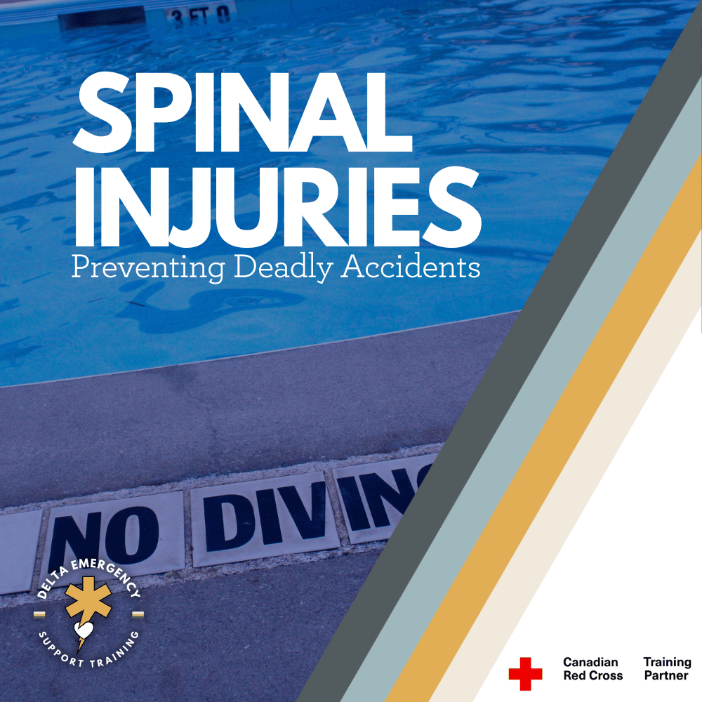
How to Prevent Spinal Injuries: Essential Steps for Safety
Spinal injuries can have devastating consequences, from paralysis to lifelong medical complications. Fortunately, many of these injuries are preventable. In this blog, we explore the top causes of spinal injuries, shocking statistics, and expert-approved safety measures to help you stay protected. Whether it’s safe driving, fall prevention, or sports safety, taking proactive steps can make all the difference. Plus, learn how Delta Emergency Support Training, a Red Cross Training Partner, provides essential first aid training to equip you with life-saving skills.
Preventing Spinal Injuries: Facts, Stats, and Essential Safety Measures
Spinal injuries are among the most severe and life-altering injuries a person can experience. Damage to the spinal cord can result in permanent paralysis, loss of sensation, and significant lifestyle changes. Understanding how these injuries occur and taking steps to prevent them is crucial for personal safety and public health.
In this blog, we will explore the causes, statistics, and preventive measures related to spinal injuries, helping you stay informed and protected.
Understanding Spinal Injuries
The spine is a complex structure made up of vertebrae, nerves, muscles, and ligaments that provide support and mobility to the body. When the spinal cord sustains damage, it can disrupt communication between the brain and the rest of the body, potentially leading to temporary or permanent disability.
Common Causes of Spinal Injuries
Spinal injuries can result from various traumatic and non-traumatic events, including:
Motor Vehicle Accidents – The leading cause of spinal cord injuries worldwide, accounting for nearly 38% of cases.
Falls – Responsible for about 32% of spinal injuries, particularly among older adults.
Sports and Recreation Accidents – Diving into shallow water, high-impact sports, and extreme activities contribute to 14% of spinal injuries.
Violence and Assaults – Gunshot wounds, stabbings, and physical attacks lead to approximately 13% of spinal cord injuries.
Medical Conditions and Diseases – Arthritis, osteoporosis, cancer, and infections can also cause spinal damage over time.
Spinal Injury Statistics
An estimated 17,900 new spinal cord injuries occur in the United States every year (National Spinal Cord Injury Statistical Center).
Males account for about 78% of spinal cord injury cases, often due to high-risk behaviors and sports activities.
The average age of a spinal injury patient has increased from 29 in the 1970s to 43 today, due to longer life expectancy and aging populations.
Lifetime costs for a spinal cord injury can range from $1.2 million to over $5 million, depending on the severity and level of paralysis.
Wearing a seatbelt and using proper vehicle restraints reduce the risk of spinal injuries in crashes by 60%.
How to Prevent Spinal Injuries
While some accidents are unavoidable, many spinal injuries can be prevented by taking proper precautions. Here are some essential strategies:
1. Safe Driving Practices
Always wear a seatbelt when driving or riding in a vehicle.
Never drive under the influence of alcohol or drugs.
Avoid distracted driving, such as texting or using a phone.
Ensure children are in proper car seats or booster seats.
2. Fall Prevention
Use handrails on stairs and install grab bars in bathrooms.
Remove tripping hazards like loose rugs and clutter.
Wear appropriate footwear with good grip.
Older adults should have regular vision check-ups and use mobility aids as needed.
3. Sports and Recreation Safety
Always wear protective gear such as helmets and padding in contact sports.
Avoid diving into shallow or unknown waters.
Use proper techniques and training for high-impact sports like football, gymnastics, and wrestling.
Follow safety rules when engaging in outdoor activities like rock climbing and skiing.
4. Workplace Safety
Use ergonomic lifting techniques to avoid back injuries.
Wear appropriate safety harnesses when working at heights.
Ensure workplaces follow OSHA safety regulations.
5. Preventing Violence-Related Injuries
Be aware of your surroundings and personal security.
Avoid high-risk areas and de-escalate confrontations when possible.
Support anti-violence initiatives and gun safety laws.
What to Do If You Suspect a Spinal Injury
If you suspect someone has a spinal injury, follow these emergency steps:
Do Not Move the Person unless they are in immediate danger.
Call 911 immediately.
Keep the Person Still by placing your hands on either side of their head to prevent movement.
Monitor Breathing and provide CPR if necessary.
Provide Comfort and Reassurance until professional help arrives.
Delta Emergency Support Training – A Red Cross Training Partner
At Delta Emergency Support Training, we provide Standard First Aid training as a Red Cross Training Partner. Our courses teach essential lifesaving skills, including how to recognize and respond to spinal injuries safely.
By taking a first aid course, you can gain hands-on experience and certified knowledge to protect yourself and others in emergencies.
Conclusion
Spinal injuries can have life-changing consequences, but many of them are preventable with proper precautions and awareness. By following safe driving habits, fall prevention strategies, and sports safety guidelines, you can significantly reduce your risk.
To be fully prepared, consider taking a first aid and injury prevention course with Delta Emergency Support Training. Being informed and trained can help you protect lives and prevent serious injuries.
How to Handle Eviscerations in Trauma: A Guide for EMRs and AFA Responders
Evisceration is a serious, life-threatening injury where internal organs protrude through an abdominal wound. As an EMR or AFA responder, it's crucial to provide immediate care by controlling bleeding, protecting exposed organs, and stabilizing the patient for transport. This blog outlines key steps and considerations to manage eviscerations effectively.
Evisceration: How EMRs and AFA Responders Should Handle Traumatic Abdominal Injuries
Evisceration is a severe and life-threatening injury that occurs when internal organs are displaced and protrude through a wound in the abdomen. As an EMR or AFA responder, managing eviscerations is critical to preventing further complications, such as infection, shock, or organ damage. Understanding how to assess and treat these injuries quickly and effectively is vital in preserving life and improving patient outcomes.
This blog will guide you through what evisceration is, how to recognize it, and how to manage the situation as a first responder.
What is Evisceration?
Evisceration refers to the protrusion of internal organs (such as the intestines or stomach) through an open wound in the abdominal area. This injury is often caused by trauma, such as:
Motor vehicle accidents
Stabbing or gunshot wounds
Blunt force trauma
Falls or industrial accidents
Unlike hernia injuries, where tissue bulges through a weak spot in the abdominal wall, evisceration involves the complete displacement of organs outside of the body. This type of injury is not only physically traumatic but also life-threatening.
Signs and Symptoms of Evisceration
The most significant sign of evisceration is the visible protrusion of internal organs through the abdominal wound. Some additional symptoms may include:
Visible intestines, stomach, or other organs outside the body.
Pain in the abdominal area.
Severe bleeding or a blood-soaked wound.
Signs of shock, such as pale skin, rapid heartbeat, and low blood pressure.
It is important to note that the presence of evisceration does not always indicate internal bleeding; however, trauma-related injuries often involve bleeding internally and externally, making it a critical condition to address.
Immediate Actions for EMRs and AFA Responders
As an EMR or AFA responder, your primary objective when managing evisceration is to prevent further harm and stabilize the patient for transport to a medical facility. Here are the key steps you should take:
1. Ensure Scene Safety
Before you begin treatment, always ensure that the scene is safe for both you and the patient. Assess any potential hazards, such as traffic, fire, or other dangerous conditions.
2. Assess Airway, Breathing, and Circulation (ABCs)
Perform a primary assessment of the patient’s airway, breathing, and circulation:
Airway: Make sure the patient’s airway is open and clear. If the patient is unconscious or struggling to breathe, perform the necessary airway interventions (e.g., head-tilt-chin-lift, suction, or intubation if trained).
Breathing: Ensure the patient is breathing effectively. If the patient is in respiratory distress, provide supplemental oxygen as needed.
Circulation: Check for signs of shock, such as weak pulse, low blood pressure, or cool, clammy skin. Severe blood loss from the abdominal organs can lead to shock, which requires immediate intervention.
3. Control Bleeding
If there is significant bleeding, it’s critical to control the hemorrhage:
Apply sterile dressings or clean cloths to the open wound. Do not attempt to push the exposed organs back into the body, as this can cause additional damage and increase the risk of infection.
Apply direct pressure around the edges of the wound to control bleeding, but do not apply pressure directly to the exposed organs.
Keep the organs moist. If sterile saline or clean water is available, dampen a sterile dressing or clean cloth and place it over the exposed organs to prevent them from drying out. Dry organs are more vulnerable to infection and damage.
4. Protect the Exposed Organs
It is crucial to protect the exposed organs from contamination, injury, and environmental elements:
Cover the organs with a sterile dressing or clean cloth moistened with sterile saline or clean water. Avoid using dry materials, which could dehydrate the organs.
Do not attempt to reinsert the organs into the body. This could cause more harm and increase the risk of further damage.
5. Position the Patient
Proper positioning is key in minimizing additional trauma:
Place the patient flat on their back (supine position) to prevent movement that could worsen the injury. Ensure the patient's head is slightly elevated if they are conscious and experiencing difficulty breathing.
If the patient is in shock, elevate their legs slightly to promote blood flow to vital organs, unless contraindicated by spinal or head injury concerns.
6. Monitor Vital Signs
Monitor the patient's vital signs closely, including pulse, blood pressure, and respiratory rate. Watch for signs of shock, such as a rapid or weak pulse, pale skin, and altered mental status. Shock is a common complication in evisceration cases, especially due to blood loss or damage to vital organs.
7. Prepare for Transport
Eviscerations require immediate transport to a hospital for surgical intervention. The patient should be rapidly transported to a trauma center or medical facility with surgical capabilities:
Alert the receiving facility about the patient’s condition, so the trauma team can be prepared for immediate action upon arrival.
Keep the patient warm to prevent hypothermia, a common complication of trauma. Use blankets or any available materials to maintain body temperature.
Complications to Watch For
Eviscerations often occur in conjunction with other serious injuries, such as:
Internal bleeding: Blood loss from damaged internal organs or blood vessels can lead to hypovolemic shock, where there is insufficient blood volume to maintain circulation.
Infection: Exposed organs are highly susceptible to infection. Keeping the wound sterile and moist is essential to minimize this risk.
Severe pain: Due to the nature of the injury, patients may experience extreme pain. If trained, provide pain relief in accordance with your scope of practice.
Conclusion
Evisceration is a traumatic and potentially fatal injury that requires quick and effective action from EMRs and AFA responders. By following the steps outlined above—controlling bleeding, protecting the exposed organs, and stabilizing the patient for transport—you can provide life-saving care and improve patient outcomes.
At Delta Emergency, our EMR and AFA training equips you with the skills needed to respond to critical injuries like evisceration. Led by experienced paramedics, we offer hands-on training to ensure you are prepared to handle any emergency situation with confidence and competence.
EMR Guide to Facial Trauma: Airway Management, Bleeding Control, and Injury Assessment
Facial trauma is a serious medical emergency that requires prompt attention. This guide for EMRs covers essential steps for managing facial injuries, including airway management, bleeding control, and fracture stabilization. Learn how to assess and treat patients with maxillofacial trauma to reduce the risk of permanent functional loss and disfigurement.
Facial Trauma - Assessment and Treatment
Tradesmen with laceration to face
Facial trauma refers to injuries involving damage to the bones or soft tissue of the face. These injuries can occur due to a variety of causes such as motor vehicle accidents, assaults (including gunshots), sports injuries, falls, chemical exposures, thermal burns, or animal bites. Because the human face is complex and contains many critical structures, such as bones, blood vessels, nerves, muscles, and sensory organs, untreated facial trauma can lead to permanent functional loss (such as problems with seeing, chewing, speaking, or swallowing) and severe disfigurement. Therefore, rapid and effective assessment and treatment are crucial in preventing long-term complications.
Facial trauma can be categorized into two primary categories: bone and soft tissue injuries. Depending on the type and severity of the trauma, the treatment and management priorities will differ. Let’s explore how to assess, treat, and what to expect when dealing with facial trauma in an emergency setting.
Key Considerations for EMRs Responding to Facial Trauma
When an EMR encounters a patient with facial trauma, the first priority is to assess the patient's airway, control bleeding, and assess for any associated injuries. This is especially critical because facial trauma can lead to airway compromise and severe bleeding, both of which are life-threatening.
Airway Management: The First Priority
One of the most critical aspects of treating facial trauma is securing the airway. The face houses the upper airway structures, and injuries to the face can easily result in obstruction. Up to 42% of patients with severe maxillofacial trauma may require intubation. If airway compromise is not addressed, it can result in hypoxia, shock, or even death.
Airway compromise in facial trauma typically occurs due to:
Soiling of the airway: Blood, vomit, or other debris can obstruct the airway, especially in patients with severe bleeding or emesis.
Obstruction: This can result from displaced soft tissue (such as the tongue), facial swelling, or foreign bodies (like dislodged teeth).
When assessing for airway compromise, EMRs should prioritize maintaining patency and anticipate potential difficulties with bag-mask ventilation due to the altered facial anatomy. For example, in Le Fort II and Le Fort III fractures, which cause significant facial displacement, it can be challenging to achieve a proper seal with a mask. If bag-mask ventilation becomes difficult, consider using a supraglottic airway device as a temporary bridge to more definitive airway management until advanced care can be provided.
Controlling Bleeding
Facial injuries, especially those involving trauma to the nose, mouth, or eyes, often result in significant bleeding. The face is highly vascular, so controlling bleeding early is vital to avoid shock and further complications. The steps to control bleeding are:
Direct Pressure: Apply direct, firm pressure over the bleeding site using sterile gauze or a clean cloth.
Elevation: If possible, elevate the patient’s head to minimize blood flow to the injury site.
Pressure Points: Use pressure points such as the carotid artery or facial artery to control hemorrhage when direct pressure is insufficient.
Hemostatic Agents: In severe cases, use hemostatic agents or dressings to assist in clotting and bleeding control.
Assessing Associated Injuries
Facial trauma often occurs as part of multi-system injuries. Once the airway is secured and bleeding is controlled, assess for other possible injuries, such as:
Head injury: Concussions, skull fractures, or intracranial hemorrhages are common in traumatic incidents that involve facial injury.
Spinal injury: Always suspect cervical spine injury in any trauma patient, especially those with altered mental status or facial fractures.
Dental injuries: Dislodged teeth or fractures to the jaw may occur in conjunction with facial trauma and may require specific management.
Eye injuries: Damage to the eyes, such as orbital fractures, corneal abrasions, or foreign bodies, should be suspected and managed accordingly.
Types of Maxillofacial Fractures
Facial fractures can be complex, and different fractures require different management strategies. Some common types of fractures include:
Le Fort Fractures:
Le Fort I: A horizontal fracture of the maxilla (upper jaw), often associated with palate and teeth displacement.
Le Fort II: A pyramidal fracture involving the maxilla, nasal bones, and orbit, which may cause significant facial deformity.
Le Fort III: A complex fracture involving the maxilla, zygomatic bones, and orbit, resulting in severe facial disfiguration and airway compromise.
Orbital Fractures: These fractures affect the bony orbit surrounding the eye. They may lead to eye injuries and double vision, requiring immediate attention to preserve vision.
Nasal Fractures: Fractures to the nasal bones are common and often associated with bleeding and airway obstruction due to the nasal passages becoming blocked with blood.
Mandible (Jaw) Fractures: Fractures of the mandible may affect the ability to chew, swallow, and speak. In some cases, the fractured jaw may obstruct the airway.
Treatment Considerations
The management of facial trauma depends on the type and severity of the injury. However, the basic principles of care involve airway management, bleeding control, and stabilization of fractures.
Airway Management
As previously mentioned, ensuring airway patency is paramount in facial trauma cases. If there is any suspicion of airway compromise, early intubation should be performed. In some cases, a supraglottic airway device may be used to bridge the gap before definitive airway management. In more severe cases, surgical airway interventions may be required.
Fracture Stabilization
Fractures, particularly those involving the mandible, maxilla, or facial bones, may need to be stabilized. The use of splints, jaw wiring, or other external fixation devices can help keep the fractures in place until surgical intervention by a plastic or maxillofacial surgeon can be performed.
Transportation
Facial trauma victims, particularly those with suspected airway compromise, require rapid transport to a hospital with appropriate resources for managing complex trauma. Transport should be done with the patient’s head elevated and airway support in place to prevent further compromise.
Conclusion
Facial trauma is a serious medical condition that requires prompt and effective intervention. As an EMR, your role in ensuring airway management, bleeding control, and assessment of associated injuries is vital in improving patient outcomes. While facial trauma may be life-threatening, the coordinated efforts of EMRs, emergency doctors, and specialists such as plastic and reconstructive surgeons can significantly reduce the risk of permanent functional loss and disfigurement. By understanding the anatomy of the face, the types of fractures, and the treatment strategies, you can provide critical care that saves lives and improves the long-term recovery of those injured in traumatic incidents.
Stay informed, stay prepared, and continue to prioritize the ABCs (Airway, Breathing, Circulation) in any trauma situation.
Ejections from Vehicles: What Every First Responder Should Know
Vehicle ejections are one of the most dangerous and traumatic incidents first responders encounter. This blog provides vital insights into the causes, injuries, and best practices for responding to ejections, as well as how advanced first aid training can help firefighters and emergency responders handle these high-risk situations effectively.
Ejections from vehicles are among the most traumatic and severe incidents that first responders—including advanced first aid professionals—encounter. These types of accidents are especially dangerous for both the ejected individual and the responders who must navigate potentially hazardous environments. In Alberta, advanced first aid training is a prerequisite for firefighters and emergency medical responders (EMRs), equipping them with the skills needed to handle these high-risk emergencies effectively.
In this blog, we’ll explore what vehicle ejections are, why they happen, the injuries that often result, and how firefighters and first responders should approach such incidents. We’ll also highlight the importance of advanced first aid training in such high-stakes situations.
What is a Vehicle Ejection?
A vehicle ejection occurs when a person is thrown from their vehicle during a crash. This can happen in a variety of ways: high-speed crashes, rollovers, or when seatbelts aren’t used or fail. Ejections are dangerous because victims often land on hard surfaces, like the road, which increases the risk of life-threatening injuries. Immediate and proper intervention is critical to give these victims the best chance of survival and recovery.
Injuries caused by ejections can range from head trauma and spinal injuries to broken bones and internal bleeding. The consequences of such injuries can be catastrophic without timely and professional care.
Causes of Ejections
Lack of Seatbelt Use: A major cause of ejections is the failure to wear seatbelts. Seatbelts significantly reduce the risk of ejection by keeping passengers securely in place during a crash.
Rollover Accidents: In rollover crashes, passengers are more likely to be ejected as the vehicle tumbles, increasing the chances of being thrown out.
High-Speed Collisions: High-speed crashes, especially when hitting stationary objects, create enough force to propel occupants out of the vehicle.
Side-Impact (T-Bone) Collisions: When a vehicle is struck from the side, particularly with high impact, the risk of ejection increases as the side windows may shatter and the force can throw an occupant from the car.
Injuries from Ejections
Ejected vehicle victims can suffer a range of injuries, many of which are life-threatening:
Head and Brain Injuries: Traumatic brain injuries (TBI) are common and can range from concussions to severe skull fractures, leading to long-term disabilities or death.
Spinal Cord Injuries: Damage to the spine can result in paralysis or death, especially if the spinal cord is compromised during the ejection.
Internal Injuries: Blunt force trauma can cause internal bleeding, ruptured organs, and significant damage to internal structures. Rapid recognition and treatment of internal injuries are essential.
Fractures and Limb Injuries: Broken limbs, ribs, and pelvis are frequent in ejected patients, especially as they come into contact with the road or surrounding objects.
Soft Tissue Injuries: Scrapes, cuts, and bruises from the force of ejection and contact with surfaces are common and can add to the patient’s overall trauma.
How to Approach Vehicle Ejections: First Responder Steps
1. Start Your Size-Up at Dispatch
The time to start assessing the situation is before you even arrive on the scene. The dispatch information provides vital clues about the nature of the crash, the type of road (residential or highway), the speed of the impact, and any hazards that may be present, like industrial vehicles or hazardous materials. This early information helps responders prepare mentally and strategically for what lies ahead.
2. Perform an Inner Circle Survey
Upon arriving at the scene, conduct a quick 360-degree assessment of the incident—what we call the "inner circle survey." Walk around the wreckage to gather information about the number of victims, vehicle conditions (engine running, fluids leaking), and possible hazards (e.g., fuel, electrical lines, airbags). Noticing these details early will help guide your approach and ensure responder safety.
Safety systems such as airbags, rollover protection devices, and seatbelt pretensioners may have deployed, which can present both advantages and hazards during rescue operations. Recognizing these features will help prevent accidental injury to responders.
3. Stabilize the Victim and the Scene
Severe Bleeding Control: Ejected victims often suffer significant blood loss due to trauma. Controlling bleeding should be your first priority. Apply direct pressure to visible wounds and use tourniquets if needed to control extremity bleeding. Hemorrhage control is vital to prevent shock and further complications.
Spinal Stabilization: Always assume spinal injury in ejected patients until proven otherwise. Quickly immobilize the head and neck using cervical collars and spinal boards, ensuring minimal movement to prevent further damage.
Treat for Shock: Victims of vehicle ejections are at a high risk for shock, due to the trauma they’ve sustained. Keep the victim warm, elevate their legs (unless there’s suspected spinal injury), and monitor their vital signs.
4. Coordinate with EMS for Transport
Once the victim has been stabilized, prepare for rapid transport. Ensure that EMS has been notified, and keep in mind that time is critical in cases involving vehicle ejections. While waiting for EMS, continue to monitor the victim and provide necessary care.
Why Advanced First Aid Is Crucial for Firefighters
In Alberta, firefighters must undergo advanced first aid training as part of their professional certification. This training is invaluable when responding to high-risk incidents like vehicle ejections, where rapid, effective care can significantly impact patient outcomes.
Advanced first aid courses equip firefighters with skills such as:
Trauma assessment and management
Spinal immobilization techniques
CPR and airway management
Hemorrhage control and shock management
Having this expertise ensures that firefighters are prepared to handle the complex and often dangerous nature of vehicle ejection incidents. The ability to recognize signs of serious injury, apply the right interventions, and work safely around hazards can be the difference between life and death for the patient.
Conclusion
Vehicle ejections are some of the most challenging and dangerous situations first responders will face. Understanding how to assess, stabilize, and transport these patients is essential for their survival and recovery. With advanced first aid training, firefighters and emergency responders are better equipped to handle the complexities of vehicle ejections, ensuring they provide the best care possible in high-pressure situations.
At Delta Emergency Support Training, we offer specialized advanced first aid courses designed to prepare firefighters, paramedics, and emergency responders for these high-stakes incidents. Our training is hands-on, scenario-based, and tailored to the specific challenges you face in the field. Reach out to us today to find out how we can help your team be prepared for anything.
About Us
At Delta Emergency Support Training, we specialize in providing advanced first aid training for firefighters and emergency responders in Alberta. Our training ensures that you have the knowledge and hands-on experience to handle complex medical emergencies, from vehicle ejections to cardiac arrest. Contact us to learn more about our training programs and how we can help you be prepared for any emergency.
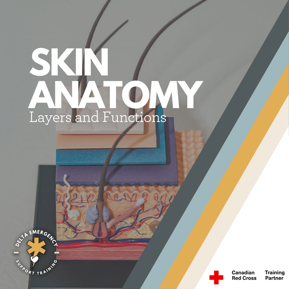
Skin Anatomy Overview for First Responders: Layers and Functions
Understanding the anatomy of the skin is essential for first responders. The skin serves as the body’s first line of defense, regulating temperature and protecting against infection. In this blog, we’ll break down the three layers of skin—epidermis, dermis, and hypodermis—and explore their functions, common injuries, and how this knowledge can help you provide better care in emergency situations.
As a first responder, you're trained to respond to a variety of emergencies, from burns and cuts to more complex trauma situations. Understanding the anatomy of the skin is crucial, as it plays a vital role in protecting the body from infection, regulating temperature, and healing injuries. In this blog, we’ll break down the layers of the skin, their functions, and how this knowledge helps you in your critical role as a first responder.
medical skin layer model
The Basics of Skin Anatomy
The skin is the body's largest organ, serving as the primary barrier between the internal systems and the outside world. It protects against physical damage, pathogens, and dehydration, while also playing a key role in temperature regulation. The skin is made up of three main layers, each with its own distinct function:
Epidermis (Outer Layer)
Dermis (Middle Layer)
Hypodermis (Subcutaneous Layer)
1. The Epidermis: The Body’s First Line of Defense
The epidermis is the outermost layer of skin, providing a waterproof barrier that shields the body from harmful substances, bacteria, and UV radiation. As a first responder, you will often encounter patients with injuries to the epidermis, such as cuts, abrasions, burns, or rashes.
Functions of the Epidermis:
Protection: The epidermis acts as the first line of defense against pathogens, chemicals, and physical damage.
Barrier to Water Loss: It helps prevent excessive loss of moisture, maintaining hydration in the body.
Skin Pigmentation: Melanin, produced in the epidermis, helps protect deeper layers from UV damage.
Sensation: The epidermis contains nerve endings that allow the skin to respond to environmental stimuli, like heat or pressure.
Important Considerations for First Responders:
Burns: Since the epidermis contains no blood vessels, burns typically affect this layer first, causing redness, swelling, and pain. In severe cases, it may also affect the dermis, leading to more serious injuries.
Infections: When the epidermis is compromised (such as in open wounds), there’s a higher risk of infection. Proper cleaning and dressing of wounds are essential to avoid bacterial infiltration.
2. The Dermis: The Skin’s Strength and Support
The dermis lies beneath the epidermis and is responsible for most of the skin's strength, elasticity, and sensation. This layer contains blood vessels, nerves, hair follicles, sweat glands, and connective tissue.
Functions of the Dermis:
Structural Support: The dermis contains collagen and elastin, proteins that provide strength and elasticity to the skin, allowing it to stretch and return to its original shape.
Temperature Regulation: Blood vessels in the dermis help regulate body temperature by constricting to conserve heat or dilating to release heat.
Sensation: The dermis is rich in nerve endings that detect pain, pressure, temperature, and touch.
Wound Healing: The dermis plays a critical role in healing skin wounds, as it contains the blood vessels and connective tissue that support tissue repair.
Important Considerations for First Responders:
Deep Cuts and Lacerations: When treating deep cuts or lacerations, you may encounter injury to the dermis, which could result in more severe bleeding due to the presence of blood vessels.
Blisters and Burns: Severe friction or thermal injuries can cause fluid buildup between the epidermis and dermis, leading to blisters. Burns can also extend into the dermis, causing damage to blood vessels and deeper tissues.
3. The Hypodermis: The Body’s Cushion and Insulator
The hypodermis, or subcutaneous layer, lies below the dermis and is primarily made of fat and connective tissue. This layer plays a significant role in protecting internal organs, insulating the body, and storing energy.
Functions of the Hypodermis:
Insulation: The fatty tissue helps regulate body temperature by providing insulation against heat loss.
Cushioning: It acts as a shock absorber, protecting bones and organs from injury by absorbing impact.
Energy Storage: The hypodermis stores fat, which serves as an energy reserve for the body.
Anchoring the Skin: It helps anchor the skin to underlying muscles and bones, giving the skin its flexibility and strength.
Important Considerations for First Responders:
Severe Trauma: In cases of deep trauma, such as blunt force injuries, the hypodermis may be damaged. This could lead to bruising or deeper tissue damage, as blood vessels in the hypodermis may rupture.
Frostbite: The hypodermis is the most affected area during frostbite, as it is the deepest layer. Severe cold temperatures can cause the fat cells to freeze, potentially leading to tissue death and the need for immediate medical intervention.
Skin Injuries and First Aid: Key Points for First Responders
As a first responder, understanding the anatomy of the skin helps you assess injuries more effectively and provide better care. Here are some common skin injuries and how they relate to the skin’s structure:
Abrasions: These involve damage to the epidermis, often caused by friction. While they are generally not severe, they can lead to infection if not properly cleaned and dressed.
Lacerations: Deep cuts can damage both the epidermis and dermis, leading to bleeding and pain. Proper wound care includes stopping the bleeding, cleaning the wound, and applying appropriate dressings.
Burns: Burns can range from superficial (affecting only the epidermis) to severe (penetrating the dermis and hypodermis). First-degree burns may cause redness and pain, while second-degree burns result in blisters and more intense pain. Third-degree burns involve damage to all layers of the skin and require immediate medical attention.
Frostbite: Exposure to extreme cold can lead to frostbite, which damages the skin, particularly the hypodermis. Treatment includes gradual rewarming and protecting the affected areas from further damage.
Infections: Skin wounds, especially when the epidermis is broken, are susceptible to infection. As a first responder, it’s critical to clean wounds thoroughly, apply sterile dressings, and monitor for signs of infection.
Conclusion: Why Skin Anatomy Matters for First Responders
The skin may seem like a simple barrier, but its structure and functions are far more complex and essential to maintaining overall health. As first responders, understanding the layers of the skin, their functions, and how injuries affect them helps you provide better care to patients. Whether you're dealing with a simple abrasion or a severe burn, this knowledge can guide your decisions on how to treat injuries quickly and effectively, reducing the risk of complications and promoting faster recovery.
By applying this understanding of skin anatomy to your daily work, you can ensure that your patients receive the best care possible in emergency situations.
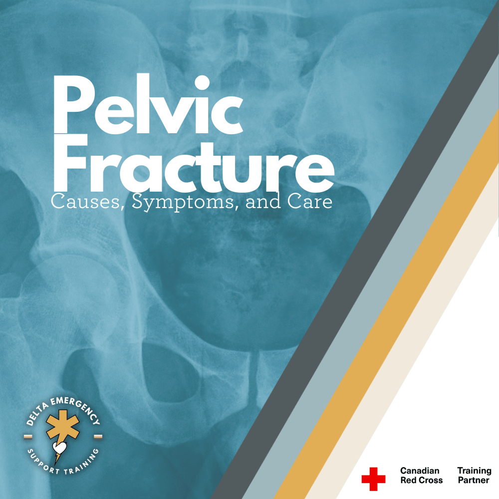
Navigating the Complexity of Broken Pelvis: Causes, Symptoms, and Care
In our comprehensive guide, we unveil the intricacies of pelvic fractures, shedding light on their causes, recognizing vital symptoms, and offering essential tips for immediate care. Unlock the secrets of this often-underestimated injury and empower yourself to make a difference in emergencies.
The pelvis, often overlooked in our daily lives, plays a pivotal role in our body's structure and stability. Yet, when a pelvic fracture occurs, it thrusts this often-neglected region into the spotlight as a site of pain, danger, and potential life-threatening complications. In this comprehensive blog, we embark on a journey to demystify pelvic fractures, exploring their origins, the subtle signs that signal their presence, and the crucial steps to provide immediate care. Join us as we uncover the intricacies of pelvic fractures, enabling you to recognize, respond to, and support those affected by this serious and often underestimated injury.
X-Ray of the pelvic region.
High-Impact Mechanisms of Injury (MOI) Leading to Pelvic Fractures
Pelvic fractures often arise from high-impact mechanisms of injury (MOI), which can deliver a devastating blow to the body's central support structure. It's essential to grasp the specific scenarios where these fractures commonly occur to take preventive measures and promptly address them. Here, we explore three prominent MOIs associated with pelvic fractures:
1. Car Accidents with Intrusion:
Car accidents, especially those involving intrusion into the passenger compartment, pose a significant risk of pelvic fractures. When a vehicle collides with another object or experiences a sudden deceleration, the forces involved can be transferred to the occupants. In such cases, the pelvis may absorb a substantial portion of the impact, leading to fractures. The use of seatbelts and properly installed airbags is crucial in reducing this risk.
2. Being Struck as a Pedestrian:
Pedestrians involved in accidents with moving vehicles face a heightened risk of pelvic fractures. When a pedestrian is struck by a vehicle, the impact forces are directed towards the pelvis, which can result in fractures. The severity of the injury depends on various factors, including the speed of the vehicle, point of impact, and the pedestrian's position. Awareness, adherence to traffic rules, and practicing safe pedestrian behaviors are essential in preventing these accidents.
3. High-Impact Sports, such as Horse Riding:
Engaging in high-impact sports like horse riding, skiing, or contact sports increases the likelihood of experiencing pelvic fractures. These activities expose individuals to abrupt movements, falls, or collisions with considerable force. In horse riding, for instance, an unexpected fall from a galloping horse can generate enough energy to fracture the pelvis. Proper safety gear, training, and adhering to safety guidelines can mitigate the risk associated with these sports.
Recognizing Indications of a Pelvic Fracture
When it comes to pelvic fractures, understanding the potential indications is paramount for swift intervention and treatment. These fractures often manifest with a combination of alarming symptoms, each serving as a red flag to signal the severity of the injury. Here, we explore some of the most common and telling indications:
1. Pelvic Instability:
One of the hallmark signs of a pelvic fracture is pelvic instability. This instability can cause a profound sense of discomfort and difficulty in maintaining balance and mobility. Individuals may experience a noticeable shift or misalignment of the pelvic bones, which can be felt when gently pressing on both sides of the pelvis. This instability can be particularly unsettling, making even simple movements a challenge.
2. Excruciating Pain:
Pelvic fractures are often accompanied by excruciating pain in the pelvic region. This pain is typically sharp, intense, and may radiate to the lower back or down the legs. The severity of the pain can vary depending on the extent and location of the fracture. It is important to note that the intensity of the pain may not correlate directly with the severity of the fracture, making it imperative to seek medical attention regardless of the perceived pain level.
3. Splayed Legs:
Another telltale sign of a pelvic fracture is the positioning of the legs. In many cases, the legs may appear splayed or twisted unnaturally due to the disruption of the pelvic bones. This visual indication can be distressing and further underscores the need for immediate medical evaluation.
4. Bruising Indicative of Internal Bleeding:
Bruising around the pelvic area can be indicative of internal bleeding, a critical concern associated with pelvic fractures. As the fractured bones can damage blood vessels within the pelvis, blood may accumulate in the surrounding tissues, causing visible bruising. This external sign of internal bleeding should not be overlooked, as it necessitates urgent medical attention.
Treatment of Pelvic Fractures
When a suspected pelvic fracture is encountered, the immediate goal is to provide stabilization and support while minimizing the risk of further injury. Proper treatment is essential to optimize outcomes and reduce complications. Here is a step-by-step guide to treating pelvic fractures:
1. Manual Stabilization:
The first and most crucial step in managing a suspected pelvic fracture is to provide manual stabilization. This involves gently supporting the pelvis with your hands to minimize movement. Care must be taken to avoid putting pressure directly on the fractured area to prevent exacerbating the injury. Manual stabilization helps in reducing pain and the risk of further damage.
2. Binding the Feet Together:
To address the splayed legs often associated with pelvic fractures and return them to a more anatomically aligned position, binding the feet together is recommended. This can be achieved by using a bandage or cloth to secure the ankles together. The goal is to maintain alignment and prevent further displacement of the fractured bones.
3. Application of a Commercial Pelvic Binder:
Commercial pelvic binders, such as the T-pod, are specially designed to provide effective stabilization for pelvic fractures. These devices are easy to apply and can help bring the fractured pelvic bones together, reducing pain and the risk of internal bleeding. Properly positioning and securing the pelvic binder is essential for its effectiveness.
4. Improvised Binding with a Sheet and Clips:
In situations where a commercial pelvic binder is not available, improvising with a sheet or any strong fabric, along with clips or other fasteners, can provide temporary stabilization. The sheet is wrapped tightly around the pelvis and secured in place to minimize movement and maintain alignment. While not as effective as commercial binders, this method can still be valuable in emergencies.
5. Administering High-Flow Oxygen:
The shock that often accompanies pelvic fractures can be life-threatening. Administering high-flow oxygen is a critical measure to help the body compensate and mitigate the effects of shock. Oxygen supplementation ensures that vital organs receive adequate oxygenation, reducing the risk of organ failure and improving the patient's chances of survival.
In Conclusion: Empowering Knowledge, Saving Lives, and Joining Delta Emergency
Our journey through pelvic fractures has highlighted the critical need for understanding this injury. Recognizing the signs, providing immediate care, and seeking prompt medical attention are paramount. Knowledge is power, and here, it can be lifesaving.
Pelvic fractures are complex, with hidden dangers such as internal bleeding and shock. Armed with this information, you're better prepared to respond effectively.
Joining Delta Emergency for your professional responder needs ensures you have access to comprehensive training, cutting-edge equipment, and a community committed to making a difference. Together, we can minimize the impact of pelvic fractures and provide the support and care that individuals deserve on their path to recovery.
Unraveling Traumatic Brain Injuries: Causes, Effects, and Signs
Unravel the complex terrain of traumatic brain injuries, where the immediate impact is just the beginning. Explore the hidden consequences as we delve into the intricate aftermath of these injuries. From internal bleeding that disrupts brain function to bruised brain tissue and the delicate damage to nerve cells, discover how hemorrhaging, contusions, and axonal injury cast a far-reaching impact. Our exploration not only sheds light on the physiological intricacies but also emphasizes the critical need for comprehensive care and healing strategies. Join us in uncovering the hidden layers of traumatic brain injuries and understanding their profound implications on both the brain and the individual's well-being.
Within the intricate realm of human health, traumatic brain injuries (TBIs) stand as complex and potentially life-altering events that can reshape the course of an individual's existence. These injuries, often stemming from accidents and unforeseen incidents, cast a broad shadow of consequences affecting cognitive, physical, and emotional well-being. In this comprehensive exploration, we delve deeply into the intricate landscape of TBIs, unearthing their causes, effects, and telltale signs. Moreover, we'll embark on an illuminating journey into the biomechanics of coup and counter coup injuries, while also delving into the internal trauma that accompanies these impactful events.
Understanding Traumatic Brain Injuries
At the heart of traumatic brain injuries lies the abrupt and forceful interaction between the brain and external forces. Such interactions lead to damage within the delicate structures of the brain, triggering a cascade of physiological and neurological disturbances. These injuries can vary in severity, ranging from the seemingly minor effects of a concussion to the profound and enduring consequences of severe brain damage.
Coup and Counter Coup: Unraveling the Mechanics
Underlying the mechanics of TBIs are phenomena known as coup and counter coup injuries. During rapid deceleration or impact, the brain can collide with the inner surfaces of the skull. This initial impact, known as coup injury, occurs at the point of contact. However, the brain's momentum doesn't cease abruptly; it continues to travel within the skull, resulting in a secondary impact on the opposite side—this is the counter coup injury. The intricate interplay between coup and counter coup impacts can lead to widespread damage and potentially severe traumatic brain injury.
Common Causes of TBI
Falls:
Everyday scenarios like falls from heights, slips on slippery surfaces, and stumbling down stairs contribute significantly to TBIs.
Motor Vehicle Accidents:
High-speed collisions, rollovers, and direct blows to the head during vehicular accidents are among the leading causes of traumatic brain injuries.
Sports-Related Injuries:
Contact sports like football, soccer, and hockey pose a notable risk of TBIs, particularly without adequate protective gear.
Assaults:
Violent incidents, physical altercations, and intentional blows to the head can culminate in traumatic brain injuries.
Signs and Symptoms of TBI
Physical Symptoms:
Common physical indicators of a TBI encompass persistent headaches, nausea, vomiting, dizziness, and sensitivity to light or noise.
Cognitive Changes:
Traumatic brain injuries frequently bring about cognitive alterations, such as confusion, memory problems, difficulty concentrating, and shifts in thinking patterns.
Emotional and Mood Alterations:
TBI sufferers often grapple with emotional shifts including irritability, mood swings, heightened anxiety, depression, and intensified emotional responses to stimuli.
Internal Hemorrhaging, Contusions, and TBI
The far-reaching consequences of TBIs often extend to internal injuries that exacerbate the damage:
Hemorrhaging:
Hemorrhaging, a serious consequence of traumatic brain injuries, emerges as a result of the forceful impact that the brain endures. The sheer intensity of the trauma can cause blood vessels within the brain to rupture, initiating internal bleeding that reverberates throughout the delicate neural terrain. This internal bleeding is not only a physical disruption but also a cascading event that disrupts the brain's intricate balance and functionality.
As blood pools within the brain, it creates pressure, a phenomenon referred to as elevated intracranial pressure. This heightened pressure exerts strain on the surrounding brain tissue, inhibiting normal circulation and potentially leading to ischemia—a lack of oxygen and nutrients for brain cells. The disrupted blood flow and accumulated blood can also trigger inflammation, exacerbating the injury's impact and hindering the brain's attempts to heal itself.
Contusions:
Contusions, akin to bruises on the brain, manifest as a direct result of the brain colliding with the unyielding bony structures of the skull. This collision leads to localized areas of brain tissue sustaining damage and becoming compromised. Similar to how a bruise forms on the skin's surface after an impact, contusions arise within the brain tissue due to the abrupt and forceful contact.
The aftermath of a contusion involves a complex interplay of cellular responses. The damaged tissue swells in response to the trauma, leading to localized swelling that can further compress adjacent brain areas. Neural pathways, responsible for transmitting signals and facilitating communication between different regions of the brain, can be disrupted due to this swelling. This disruption can lead to impairments in cognitive, sensory, and motor functions, depending on the location of the contusion.
Axonal Injury:
The swift movement of the brain within the confines of the skull can result in a phenomenon known as axonal injury—a condition where nerve cells, or axons, endure stretching, twisting, or even shearing. These axons serve as the communication highways of the nervous system, transmitting electrical impulses that enable various bodily functions and cognitive processes.
In cases of axonal injury, these vital communication pathways become compromised. The structural integrity of the axons is disrupted, leading to a range of neurological impairments. Depending on the severity of the injury and the extent of axonal damage, individuals may experience motor deficits, cognitive dysfunction, and sensory disturbances. The complexities of axonal injuries are further compounded by the fact that these microscopic structures are challenging to repair, necessitating innovative approaches to encourage nerve cell regeneration and functional recovery.
Seeking Medical Attention and the Road to Recovery
Recognizing the signs of a TBI and swiftly seeking professional medical attention are pivotal steps in the journey toward effective diagnosis and treatment. The path to recovery from a traumatic brain injury is as diverse as the injuries themselves. Factors such as the severity of the injury and the effectiveness of medical interventions all contribute to the healing process. Rehabilitation therapies, cognitive exercises, and supportive care all play a pivotal role in aiding recovery and restoring quality of life.
Conclusion
Traumatic brain injuries stand as complex events with multifaceted implications for both the individual and their support network. By cultivating a deep understanding of the causes, effects, and signs of TBIs, we are empowered to advocate for enhanced safety measures and expedient medical intervention. The intricate biomechanics of coup and counter coup injuries underscore the dynamic relationship between the brain and the protective skull. As we strive for safer environments and heightened awareness, we take significant strides in diminishing the impact of TBIs on lives, while fostering a culture of brain health and holistic well-being.

