The Delta Dispatch

CFD Application Guide for Aspiring Firefighters

Dreaming of becoming a Calgary firefighter? This guide walks you through the CFD application process, including essential certifications, physical fitness tips, suggested courses, and how Delta Emergency Support Training can help you succeed.

If you’ve ever dreamed of wearing the Calgary Fire Department uniform, you’re not alone. Every year, countless motivated individuals from all walks of life apply to join one of Canada’s most respected firefighting teams. Whether you’re coming off the rigs, working in the trades, or ready for a meaningful career that serves your community, the CFD recruitment process is both challenging and rewarding.

At Delta Emergency Support Training, we’ve helped many students prepare for that next step, and several of our instructors are currently serving with CFD. So, if you’re serious about applying, here’s what you need to know about the process and how to set yourself up for success.

Step 1: Meeting the Basic Requirements

Before applying, make sure you meet the minimum qualifications. Applicants must:

  • Be at least 18 years old

  • Be legally entitled to work in Canada

  • Have a high school diploma or equivalent

  • Hold a valid Class 5 driver’s licence (with a clean record)

Medical fitness is crucial — you’ll need to pass a comprehensive medical assessment that ensures you can safely perform the physical demands of the job. Strong cardiovascular health, vision, and hearing are essential for operational readiness.

Step 2: Educational and Certification Requirements

To apply for CFD, you’ll need a few essential certifications. One of the most important is Advanced First Aid (AFA), which you can earn through Delta Emergency Support Training.

Our AFA course provides the hands-on medical foundation you’ll rely on every day as a firefighter — from patient assessment and airway management to emergency scene care. We’ve even built rotation-friendly schedules for those still working in camp or on the rigs, so you don’t have to put your career on hold to train.

It’s important to note that CFD does not require applicants to hold NFPA 1001 Level I and II firefighter training, which is often mandatory for many other departments across Canada. This makes CFD an ideal opportunity for motivated individuals looking to make the leap into firefighting without already having that certification.

Step 3: The Application and Testing Process

The CFD recruitment process typically includes several stages:

  • Online Application: Submit your resume, certifications, and supporting documentation.

  • Written Exam: Designed to assess problem-solving, mechanical reasoning, and situational judgment.

  • Physical Fitness Test: A demanding, firefighter-specific test that measures endurance, strength, and agility.

  • Interview: You’ll discuss your motivations, teamwork experience, and understanding of community service.

  • Background Check: Ensures candidates uphold the integrity and professionalism expected of CFD members.

The process is competitive, but with the right preparation and attitude, it’s absolutely achievable.

Step 4: How to Stand Out as an Applicant

Want to stand out in the hiring process? Focus on building the qualities that CFD values most: teamwork, communication, leadership, and resilience.

  • Physical Conditioning: Prioritize strength, endurance, and functional fitness — firefighting is demanding work.

  • Community Involvement: Volunteer with local emergency services, community programs, or charities. It demonstrates commitment and service-mindedness.

  • Communication and Leadership: Practice staying calm under pressure and leading effectively in high-stress situations.

CFD is looking for individuals who bring heart, reliability, and the willingness to learn — not just technical skill.

Step 5: Suggested Courses to Strengthen Your Resume

While CFD doesn’t require NFPA 1001 firefighter certification, there are plenty of valuable courses that can give you a competitive edge and help you hit the ground running once you’re accepted.

Consider training in areas such as:

  • Confined Space Awareness and Rescue – Learn safe entry, monitoring, and rescue techniques.

  • Rope Rescue (Basic & Advanced) – Develop technical rope-handling skills used in high-angle and low-angle rescues.

  • Hazardous Materials Awareness (HAZMAT) – Understand how to recognize and safely respond to dangerous goods incidents.

  • Transportation of Dangerous Goods (TDG) – An important credential for anyone working around hazardous substances.

  • ICS (Incident Command System) Training – Build your understanding of emergency scene management and structure.

Many of these courses are available through technical safety providers or emergency training academies — and they all show CFD that you take professional development seriously.

Step 6: Training and Career Development

Once selected, recruits undergo an intensive 16-week CFD Fire Training Academy program, covering everything from fire suppression and rescue techniques to hazardous materials response and medical care.

Graduation isn’t the end of learning — CFD firefighters continue developing through specialized courses, leadership tracks, and ongoing professional education.

How Delta Can Help You Get There

We know what it takes to succeed in the CFD process because our team has been there. At Delta Emergency Support Training, we’ve helped countless students earn their Advanced First Aid certification and gain the skills and confidence they need to take the next step.

Several of our instructors are active CFD members, offering insider guidance, mentorship, and firsthand insights into the realities of the job. Many of our graduates have successfully transitioned from oilfield work or trades into full-time firefighting careers — proving it’s never too late to make the change.

Your Firefighter Journey Starts Here

Applying for the Calgary Fire Department is a big step, but it’s one that can lead to an incredible career of purpose, teamwork, and community impact.

If you’re ready to make a change — to trade in your rotation schedule for a fire hall shift and a new sense of purpose — start today.

At Delta Emergency Support Training, we offer Advanced First Aid courses, including camp rotation-friendly schedules that fit your lifestyle. You bring the dedication — we’ll help you build the foundation.

Read More
Advanced First Aid, IV, Fluid, Pre-hospital, ITLS, EMR, Firefighter Kumari Malhotra Advanced First Aid, IV, Fluid, Pre-hospital, ITLS, EMR, Firefighter Kumari Malhotra

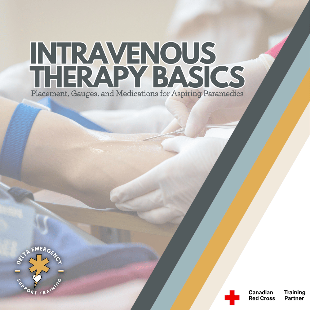
Intravenous Therapy for Beginners: Placement, Gauges & Medications

Intravenous (IV) therapy is a key skill for future paramedics and advanced responders. This beginner’s guide covers vein selection, catheter sizes, and common medications, helping you prepare for PCP training and real-world prehospital care.

For many first responders, advancing from Advanced First Aid or Emergency Medical Responder certification to Primary Care Paramedic training is an exciting milestone. It’s where your skills evolve from basic emergency management to a more advanced approach to prehospital medical care.

One of the essential new skills taught in PCP programs is intravenous (IV) therapy, a procedure that allows responders to deliver fluids and medications directly into the bloodstream for rapid, controlled treatment.

While IV initiation is not within the EMR or AFA scope of practice, understanding what it involves — and how it supports patient care — gives you a valuable head start.

What Is IV Therapy?

IV therapy involves inserting a sterile catheter into a patient’s vein to establish direct access to the circulatory system. This line can then be used to deliver fluids, medications, or draw blood samples. IV access is one of the fastest, most reliable ways to stabilize a patient in critical or emergent situations.

When Are IVs Needed?

Paramedics and advanced responders use IVs in a wide range of medical and trauma calls, such as:

  • Fluid resuscitation: Treating dehydration, burns, or blood loss

  • Medication delivery: For cardiac, allergic, or diabetic emergencies

  • Pain or nausea management: In controlled, monitored settings

  • Cardiac arrest care: To administer life-saving drugs quickly

  • Trauma management: When oral or intramuscular routes aren’t viable

Learning to Start an IV in PCP School

During PCP training, students learn the anatomy of veins, infection control, and catheterization techniques through classroom instruction, practice labs, and clinical placements. You’ll first work on training arms to master vein palpation, needle handling, and aseptic technique before performing supervised IV starts in clinical settings.

The process includes:

  1. Gathering and preparing sterile equipment

  2. Identifying a suitable vein

  3. Cleansing the site

  4. Inserting the catheter bevel-up

  5. Confirming flashback and advancing the catheter

  6. Securing the line and beginning the infusion

Common IV Insertion Sites (and Why They’re Chosen)

Vein selection is critical. Choosing the right site helps reduce complications, ensures proper flow, and keeps the patient comfortable.

Here are the most common IV entry sites used in prehospital and clinical settings:

1. Dorsal Hand Veins

These include the cephalic, basilic, and dorsal metacarpal veins on the back of the hand.

  • Pros: Easy to visualize and access; great for smaller catheters.

  • Cons: More sensitive; can be uncomfortable during movement or fluid administration.

  • Common Uses: General medication or fluid delivery when other veins are less accessible.

2. Forearm Veins

The cephalic and basilic veins along the forearm are often the first choice for IV starts.

  • Pros: Straight path, good flow rate, and easier stabilization with tape.

  • Cons: May roll or collapse if dehydrated; limited space for reattempts.

  • Common Uses: General medical and trauma patients, fluid resuscitation, medication administration.

3. Antecubital Fossa (Inner Elbow)

The median cubital, cephalic, and basilic veins in the crook of the arm are popular for rapid access.

  • Pros: Large veins, high flow rates — ideal for trauma or blood draws.

  • Cons: Restricts arm movement; prone to kinking if patient bends the elbow.

  • Common Uses: Emergency IV access, large-bore lines, or when rapid administration is needed.

4. External Jugular Vein (Neck)

Used in some prehospital or critical care settings when peripheral access isn’t possible.

  • Pros: Easy to locate in patients with difficult peripheral veins; high flow.

  • Cons: Requires skill and patient cooperation; greater risk if improperly managed.

  • Common Uses: Trauma, shock, cardiac arrest, or situations where arm veins are inaccessible.

5. Lower Extremity Veins (Dorsal Foot or Great Saphenous)

Used mainly when upper extremities are unavailable.

  • Pros: Alternative access in infants or patients with poor upper body veins.

  • Cons: Slower flow, higher infection risk, limited mobility.

  • Common Uses: Pediatrics, last-resort adult IV access.

Understanding IV Gauge Sizes

The gauge of an IV catheter refers to its diameter. Smaller numbers mean larger catheters.

  • 14–16G: For trauma or massive fluid resuscitation

  • 18G: For blood administration or major fluid replacement

  • 20G: General adult use; medications and moderate fluid therapy

  • 22–24G: Pediatrics, elderly, or fragile veins

Gauge choice depends on patient size, vein condition, and the urgency of the situation.

Common IV Medications in Paramedic Practice

While specific drug use depends on provincial protocols, IV therapy often supports:

  • Normal Saline (0.9% NaCl) – Fluid replacement or line maintenance

  • Dextrose (D10, D50) – Hypoglycemia management

  • Epinephrine – Cardiac arrest or anaphylaxis (specific concentrations)

  • Morphine / Fentanyl – Pain management (under standing orders)

  • Ondansetron – Nausea control

  • Nitroglycerin (IV form) – Chest pain management (advanced protocols)

Each medication requires strict dosage control, monitoring, and adherence to medical direction — something you’ll master in paramedic pharmacology.

Why IV Skills Matter

For professional responders, starting an IV isn’t just about inserting a catheter — it’s about stabilizing patients efficiently, safely, and confidently. In trauma, shock, or medical crises, IV access can mean the difference between life and death.

If you’re currently an EMR or AFA graduate considering the PCP path, familiarizing yourself with IV principles now will make your transition smoother and your training easier.

Final Thoughts

Learning to start an IV marks a major step forward in your responder journey. Though outside the AFA and EMR scope, it’s a skill that defines advanced prehospital care. As you move toward your Primary Care Paramedic program, understanding IV therapy — from vein anatomy to gauge selection — gives you a solid foundation for success.

Read More
Advanced First Aid, BLS, Firefighter Kumari Malhotra Advanced First Aid, BLS, Firefighter Kumari Malhotra

From the Rigs to the Fire Hall: Get Your Advanced First Aid Certification in 2026

Thinking about leaving the rigs to pursue firefighting? Our Camp Rotation Advanced First Aid courses for 2026 are designed for rig workers with demanding schedules. Get certified, gain real-world emergency skills, and take the first step toward a career saving lives.

You’ve worked the rigs, long shifts, tough conditions, and no room for hesitation. Now you’re ready for the next challenge: a career in firefighting or emergency response. The skills you’ve built on the rigs — teamwork, problem-solving, and staying cool under pressure — are exactly what fire departments and responder agencies are looking for. All you need is the right certification to start that journey.

That’s where we come in. Our Advanced First Aid (AFA) program is designed to bridge the gap between industrial safety and professional emergency response. You’ll learn how to manage trauma, perform advanced assessments, and provide life-saving care — all skills that will set you apart when applying to firefighter programs or first responder roles.

And because we know your schedule doesn’t stop for training, we’ve launched NEW Camp Rotation AFA Schedules for 2026! Whether you’re on a 2-week rotation or transitioning out of camp work, our courses are built to fit around your shifts — so you can train without losing time on the job.

By earning your AFA certification, you’ll not only meet entry requirements for many emergency services programs, but also gain the confidence and medical readiness expected in the field.

Camp worker on a 2-week rotation? Check out our NEW Camp Rotation AFA Schedules and start preparing for your next chapter — from the rigs to the fire hall.

👉 Register today and take the first step toward a career that puts your experience and drive to work saving lives.

Read More

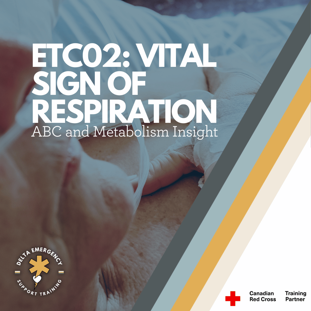
Rate, Rhythm, and Quality: How to Assess Pulse and Breathing in the Field

Assessing rate, rhythm, and quality of pulse and respirations is essential for identifying serious patient conditions. Learn how to conduct these key evaluations in the field with accuracy.

When assessing a patient, rate, rhythm, and quality are more than routine checklist items — they’re vital indicators of what’s happening beneath the surface. Whether you’re an Advanced First Aider, EMR, or future paramedic, mastering these three elements helps you recognize subtle signs of deterioration before they become life-threatening.

🩺 Assessing the Pulse

1. Rate

The rate refers to how many times the heart beats per minute (bpm).

  • Adult: 60–100 bpm

  • Child: 80–100 bpm

  • Infant: 100–160 bpm

Tachycardia (a rate above normal) may indicate fever, shock, pain, dehydration, or stress.
Bradycardia (a rate below normal) may appear in trained athletes, hypothermia, or head injuries affecting brain function.

👉 Field Tip: Always consider the context — a runner at rest with a pulse of 52 bpm may be fine, while a trauma patient with the same rate might be in serious trouble.

2. Rhythm

Rhythm describes whether the beats occur at regular intervals.

  • Regular rhythm: Evenly spaced beats (normal sinus rhythm).

  • Irregular rhythm: Uneven or skipping beats (possible arrhythmia).

Listen for patterns like regularly irregular or irregularly irregular, which may indicate cardiac abnormalities.

👉 Field Tip: Use your watch’s second hand or a digital timer — count for a full 60 seconds if rhythm appears irregular.

3. Quality

The quality of a pulse tells you about strength and perfusion.

  • Strong / bounding: Common in fever or hypertension.

  • Weak / thready: Seen in shock, blood loss, or dehydration.

  • Absent: Immediate life threat — initiate advanced interventions.

👉 Field Tip: Compare central (carotid) and peripheral (radial) pulses. A weak or absent radial pulse may signal dropping blood pressure or poor perfusion.

🌬️ Assessing Respirations

Just like the pulse, respirations are assessed for rate, rhythm, and quality.

1. Rate

Normal respiratory rates:

  • Adult: 12–20 breaths per minute

  • Child: 20–30 bpm

  • Infant: 30–60 bpm

Tachypnea (rapid breathing) often indicates hypoxia, shock, or anxiety.
Bradypnea (slow breathing) can suggest head injury, narcotic overdose, or severe illness.

👉 Field Tip: Always watch the chest discreetly, so patients don’t alter their breathing pattern.

2. Rhythm

Normal respirations are smooth and regular.

  • Irregular rhythm: May indicate neurological damage, fatigue, or shock.

  • Cheyne-Stokes respirations: Periods of deep breathing alternating with apnea — often seen in brain injury or end-of-life situations.

  • Agonal gasps: Ineffective, irregular breaths seen in cardiac arrest.

👉 Field Tip: If the rhythm looks unusual, describe exactly what you see — “deep, gasping, irregular respirations” gives dispatchers or incoming paramedics more useful information than just “irregular.”

3. Quality

Respiratory quality reflects effort, depth, and sound.

  • Normal: Quiet and effortless.

  • Shallow: May indicate pain, shock, or fatigue.

  • Labored: Look for use of accessory muscles, nasal flaring, or grunting.

  • Noisy: Listen for wheezing (airway constriction), gurgling (fluid), or stridor (upper airway obstruction).

👉 Field Tip: Note any cyanosis (bluish lips or fingertips), which can signal inadequate oxygenation.

🚑 Putting It All Together: Why It Matters

Rate, rhythm, and quality are part of every primary and secondary survey. These details guide your priorities:

  • Recognizing shock early by weak, rapid pulse and shallow respirations.

  • Identifying hypoxia through tachypnea or labored breathing.

  • Catching cardiac irregularities that could indicate impending arrest.

In short, these three simple checks — rate, rhythm, and quality — can reveal complex physiological problems long before advanced tools do.

💡 Key Takeaways for First Responders

  • Always compare findings to baseline vitals and the patient’s overall presentation.

  • Document what you observe, not just normal/abnormal.

  • Repeat assessments frequently — deterioration can happen quickly.

  • Communicate trends to higher-level care providers for continuity.

Final Thought

Assessing rate, rhythm, and quality isn’t about memorizing numbers — it’s about seeing the patient behind the vitals.The best responders know that a weak, irregular pulse or labored breathing tells a story. Learn to listen carefully — it could be the difference between early intervention and missed warning signs.

Read More

Ejection Trauma: What First Responders Need to Know About High-Impact MVCs

Ejection trauma is one of the most catastrophic outcomes of a motor vehicle collision. For first responders, understanding how to assess, stabilize, and prioritize care for these high-impact patients is critical. Here’s how to stay calm, organized, and effective in the moments that matter most.

When a person is ejected from a vehicle during a crash, the outcome is almost always severe. Ejection trauma represents one of the most catastrophic mechanisms of injury emergency responders will encounter. Whether partial or complete, being thrown from a vehicle amplifies the forces acting on the body—turning an already violent impact into a multi-system trauma event.

What Is Ejection Trauma?

Ejection trauma occurs when a vehicle occupant is completely or partially thrown out of the vehicle during a collision. This typically happens when seat belts are not worn or fail, or when the force of impact breaches the vehicle’s structure.

There are two main types:

  • Partial ejection – The patient is partially expelled, often trapped by the seat belt or vehicle frame, resulting in crushing or shearing injuries.

  • Complete ejection – The patient is fully thrown clear of the vehicle, often resulting in secondary impacts with the ground or other objects.

Mechanism of Injury (MOI)

Ejection trauma is classified as a high-mechanism MOI, and responders should immediately suspect:

  • Severe blunt trauma

  • Multiple fractures

  • Spinal, head, and internal injuries

  • Airway compromise or shock

The energy transfer doesn’t stop at the first impact—the patient often experiences multiple points of contact:

  1. The initial crash inside the vehicle

  2. Contact with the vehicle structure on exit

  3. Impact with the ground or another surface

  4. Potential secondary impact with objects (trees, poles, other vehicles)

Each of these contributes to a complex and unpredictable injury pattern.

Common Injuries Associated with Ejection

Responders should anticipate multi-system trauma, including:

  • Head and neck injuries: Concussions, skull fractures, traumatic brain injuries (TBI)

  • Spinal trauma: High risk of spinal fractures or cord damage due to rotational forces

  • Thoracic injuries: Flail chest, pneumothorax, hemothorax, cardiac contusions

  • Abdominal trauma: Liver or spleen lacerations, internal bleeding

  • Orthopedic injuries: Long bone fractures, pelvic fractures, amputations

  • Soft tissue injuries: Severe abrasions, degloving injuries, lacerations

Scene Assessment

When responding to an MVC with possible ejection, scene safety and situational awareness come first.

  • Scan the entire area—patients may be thrown significant distances or hidden in brush, ditches, or behind obstacles.

  • Note patient position relative to the vehicle; it offers key clues about trajectory and energy transfer.

  • Look for additional hazards: leaking fuel, oncoming traffic, downed power lines.

Patient Management in the Field

Ejected patients require rapid, systematic assessment and early activation of advanced care.

1. Scene Safety & Triage

  • Request additional units and air transport early.

  • Perform a global scan for multiple patients.

2. Primary Survey (Check, Call, Care)

  • Airway: Suspect spinal injury—manually stabilize head and neck, use jaw-thrust if needed.

  • Breathing: Expose the chest, check for symmetry and adequate air movement.

  • Circulation: Control major bleeding immediately. Prioritize rapid identification of life-threatening hemorrhage.

3. Secondary Survey

  • Head-to-toe assessment, noting deformities, contusions, abrasions, punctures, burns, tenderness, lacerations, and swelling (DCAP-BTLS).

  • Monitor vitals frequently—changes can indicate internal bleeding or decompensating shock.

  • Maintain spinal motion restriction (SMR) if indicated.

4. Shock Management

  • Treat for hypovolemia: position appropriately, keep warm, and prepare for rapid transport.

  • Oxygen administration and airway management should be prioritized.

The Role of Prehospital Responders

As a first responder, your calm and methodical approach can make all the difference. Recognizing the severity of ejection trauma early allows for:

  • Prioritizing rapid extrication and transport

  • Effective communication with dispatch and incoming EMS

  • Early notification of trauma centers

Ejection = Severe Trauma Until Proven Otherwise.
Never underestimate the forces involved—patients may appear stable initially but can deteriorate quickly.

Key Takeaways

  • Ejection trauma is one of the deadliest outcomes of an MVC.

  • Always assume multi-system trauma and spinal involvement.

  • Focus on airway, breathing, and circulation while minimizing movement.

  • Rapid transport and early hospital notification are essential.

  • Scene safety, teamwork, and clear communication are critical under pressure.

Final Thoughts

Every ejection call reminds responders why scene assessment and mechanism recognition matter so much. Behind every statistic is a human life—one that depends on the calm, structured, and skilled actions of those first on scene. Whether you’re an Advanced First Aider, EMR, or paramedic student, understanding ejection trauma prepares you to manage chaos with confidence and compassion.

Read More
Advanced First Aid, BLS, BLS CPR, Cardiac Kumari Malhotra Advanced First Aid, BLS, BLS CPR, Cardiac Kumari Malhotra

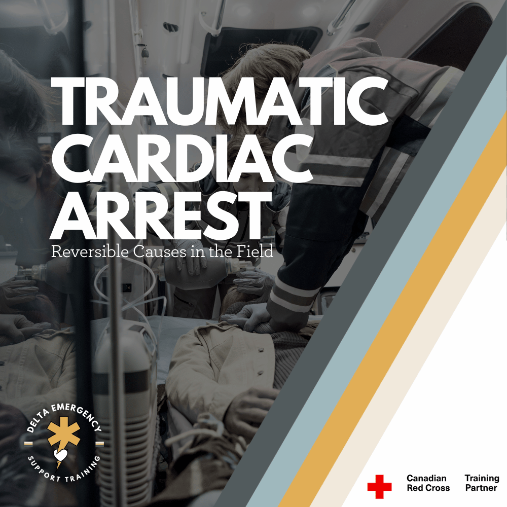
Traumatic Cardiac Arrest: What Every Professional Responder Should Know

Traumatic cardiac arrest is one of the most demanding situations a responder can face. This in-depth guide explains how to rapidly identify reversible causes, manage airway and bleeding, and provide effective field care to improve survival chances in severe trauma cases.

Traumatic cardiac arrest (TCA) represents one of the most challenging situations in prehospital care. Outcomes are often poor, but when managed rapidly and effectively, survival with good neurological recovery is possible. This blog explores what TCA is, its causes, assessment and treatment priorities, and best practices for professional responders in Canada.

What Is Traumatic Cardiac Arrest?

A traumatic cardiac arrest occurs when a patient’s heart stops as a direct result of major trauma. This trauma can be blunt (such as falls or motor vehicle collisions) or penetrating (such as gunshots or stab wounds).

Unlike medical cardiac arrests, TCA often stems from reversible physical causes rather than primary heart dysfunction. The most common reversible causes include:

  • Severe hemorrhage (uncontrolled bleeding)

  • Hypovolemia (loss of circulating volume)

  • Hypoxia (insufficient oxygen supply)

  • Tension pneumothorax (collapsed lung causing pressure on the heart)

  • Cardiac tamponade (blood filling the pericardial sac and compressing the heart)

How Common Is TCA and What Are the Survival Trends?

Traumatic cardiac arrests are less common than medical arrests but are often more complex. Historically, survival rates have been extremely low—typically below 2%. However, in cases where the underlying cause is rapidly identified and corrected, patients can and do survive, especially when there are early signs of life or immediate advanced care.

Outcomes tend to be more favorable in penetrating trauma compared to blunt trauma, largely due to the speed at which reversible causes can be treated.

Key Priorities in Managing TCA

When seconds matter, responders must focus on interventions that directly address reversible causes. The following priorities guide professional care:

PriorityActionPurposeControl Hemorrhage / HypovolemiaApply direct pressure, tourniquets, wound packing, or pelvic binders.Prevent further blood loss and restore perfusion.Airway and OxygenationClear the airway and ventilate effectively. Use supraglottic or advanced airways if trained and authorized.Correct hypoxia, one of the leading causes of TCA.Relieve Mechanical ObstructionsPerform needle decompression for tension pneumothorax or use a chest seal for open chest wounds.Restore normal breathing and heart function.High-Quality CPROnly once reversible causes are being managed. Focus on effective compressions with minimal interruptions.Maintain perfusion if reversible causes have been addressed.Rapid TransportTransport quickly to a trauma centre if signs of life are present.Definitive care is essential for survival.

Trauma Type and Approach

Penetrating Trauma

Penetrating injuries, such as gunshots or stabbings, can cause localized damage but often have a shorter timeline to intervention. Quick chest decompression, bleeding control, and airway management can make a significant difference in survival outcomes.

Blunt Trauma

Blunt trauma, such as from high-impact collisions or falls, often causes widespread internal injuries and massive bleeding. Survival rates are generally lower, and early recognition of futility is crucial when there are no signs of life and prolonged downtime.

Canadian Best Practices and Field Considerations

Canadian EMS and paramedic protocols emphasize a systematic, cause-driven approach to TCA management:

  • Address all reversible causes simultaneously.
    Control bleeding, relieve pneumothorax, and manage airway without delay.

  • Obtain IV/IO access and administer fluids as indicated for hypovolemia.

  • Perform bilateral needle decompressions if chest trauma is suspected.

  • Apply pelvic binders when pelvic fractures or major lower-body trauma are likely.

  • Minimize scene time. Rapid transport to a trauma centre remains a priority.

  • Epinephrine is often deprioritized, as it offers limited benefit when the heart has stopped due to physical injury rather than electrical failure.

  • Termination of Resuscitation (TOR) may be considered if there are no signs of life, asystole, and extended downtime with no reversible cause found.

Common Challenges for Responders

Traumatic cardiac arrests demand rapid thinking and precise teamwork. Common challenges include:

  • Identifying reversible vs. irreversible causes. It can be difficult to determine whether the arrest is trauma-induced or secondary to a medical event.

  • Time pressure. Every second counts, especially when massive hemorrhage or tension pneumothorax is suspected.

  • Limited prehospital tools. Not all regions permit advanced procedures like thoracotomy or pericardiocentesis.

  • Balancing interventions. CPR may worsen bleeding if hemorrhage is uncontrolled, so priorities must be reassessed continually.

  • Transport logistics. Long transport times may influence whether resuscitation efforts continue.

Step-By-Step Approach to Traumatic Cardiac Arrest

  1. Ensure scene safety and don PPE.

  2. Rapidly assess mechanism and timeline (blunt vs. penetrating, time of arrest, signs of life).

  3. Control massive bleeding immediately. Use direct pressure, tourniquets, or wound packing.

  4. Manage the airway and ventilate. Provide oxygen and ensure adequate chest rise.

  5. Relieve chest pressure. Perform needle decompression and apply chest seals as indicated.

  6. Provide fluid resuscitation. Administer IV or IO fluids per protocol.

  7. Perform CPR if appropriate. Begin once reversible causes are actively addressed.

  8. Transport rapidly to a trauma facility if ROSC occurs or if the patient shows any signs of life.

  9. Consider termination if no response and all reversible causes have been managed.

Why TCA Differs from Medical Cardiac Arrest

In medical cardiac arrest, the primary issue is usually electrical or metabolic failure of the heart. In TCA, the heart often stops because of physical factors such as massive bleeding or chest injury. As a result, standard interventions like defibrillation or epinephrine are secondary to correcting the underlying mechanical or circulatory issues.

In short — treat the cause, not just the rhythm.

Realistic Scenarios

  • Penetrating Chest Trauma: A patient with a gunshot wound to the chest is pulseless on arrival. Responders immediately apply a chest seal, decompress the chest, manage the airway, and control external bleeding.

  • Blunt Pelvic Trauma: Following a vehicle collision, the patient has severe pelvic instability and no pulse. A pelvic binder is applied, external bleeding controlled, airway secured, and fluids initiated during transport.

Final Thoughts

Traumatic cardiac arrest demands composure, coordination, and deep understanding of trauma physiology. While survival rates remain low, outcomes improve significantly when responders rapidly identify and correct reversible causes.

Every second counts — and with the right interventions, professional responders can give trauma patients their best chance at survival.

Read More
Advanced First Aid, EMR, First Responder Kumari Malhotra Advanced First Aid, EMR, First Responder Kumari Malhotra

Treating Gunshot Wounds: A Professional Responder’s Guide

Gunshot wounds are life-threatening emergencies that require quick, precise action. This guide for professional responders covers wound assessment, bleeding control, chest seal application for thoracic injuries, and key steps for safe transport to trauma centers.

Gunshot wounds (GSWs) are among the most critical emergencies a responder can encounter. Severity depends on bullet type, trajectory, and location, making rapid assessment and precise intervention essential. Professional responders must be prepared to manage everything from external bleeding to life-threatening internal injuries.

Gunshot Wounds in Canada: Context & Stats

Understanding the prevalence and patterns of firearm injuries in Canada provides important context:

  • Between 2016–2020, Canada saw over 4,000 deaths and thousands of hospitalizations due to firearm injuries.

  • Fatal injuries are most often suicides (~72%), followed by homicides (~23%), with unintentional injuries and law enforcement incidents making up smaller percentages.

  • Non-fatal injuries frequently result from assault or accidental shootings, highlighting the need for skilled first responders.

  • Young adult males (20–34) are most commonly affected, emphasizing the importance of rapid intervention in high-risk scenarios.

Scene Safety & Initial Approach

Before approaching the patient:

  1. Ensure scene safety. Gunshot incidents may occur in violent settings—confirm the area is secure and involve law enforcement if necessary.

  2. Use proper PPE. Gloves, eye protection, and body protection may be required depending on the scene.

  3. Triage and rapid assessment. Identify life-threatening injuries first, especially massive bleeding or airway compromise.

Primary Survey: C-ABCDE Approach

Professional responders often use a C-ABCDE approach, prioritizing massive bleeding first:

  • C – Massive Hemorrhage: Apply direct pressure, hemostatic dressings, or tourniquets (for limbs). Junctional areas may require packing or specialized devices.

  • A – Airway: Ensure airway is clear; use adjuncts if necessary. Be alert for obstructions from blood, tissue, or debris.

  • B – Breathing: Evaluate for chest injuries. Gunshot wounds to the thorax can cause pneumothorax, tension pneumothorax, or hemothorax.

  • C – Circulation: Monitor vitals, assess for shock, establish IV access if in scope, and be ready to administer fluids per protocol.

  • D – Disability: Evaluate neurological status (AVPU or GCS) and look for signs of head or spinal injury.

  • E – Exposure & Environment: Fully assess injuries while keeping the patient warm and preserving dignity.

Treating Gunshot Wounds by Location

Extremities (Arms & Legs)

  • Risks: Severe bleeding, fractures, nerve damage.

  • Intervention: Direct pressure, tourniquets for limbs, immobilize fractures, monitor for shock.

Junctional Areas (Groin, Axilla, Neck)

  • Risks: Major vessel injury; uncontrolled bleeding.

  • Intervention: Use pressure and hemostatic dressings; junctional tourniquet if available.

Chest

Chest injuries are especially dangerous due to risk of lung collapse, cardiac injury, or massive internal bleeding.

  • Signs: Difficulty breathing, sucking chest wound, rapid deterioration, shock.

  • Intervention:

    1. Chest Seal: Apply to open chest wounds to prevent air entry and reduce risk of tension pneumothorax. Venting seals allow trapped air to escape.

    2. Oxygen Support: Provide high-flow oxygen if trained.

    3. Positioning: Keep the patient upright if comfortable to assist breathing.

    4. Rapid Transport: Patients with chest GSWs require urgent trauma care.

Abdomen & Pelvis

  • Risks: Internal organ damage, significant bleeding.

  • Intervention: Cover exposed organs with sterile, moist dressings, avoid manipulation, monitor vitals, rapid transport.

Head & Neck

  • Risks: Airway compromise, brain injury, severe bleeding.

  • Intervention: Protect cervical spine, manage airway, control bleeding, rapid transport.

Special Considerations

  • Bullet Energy & Cavitation: High-velocity bullets create more tissue damage due to cavitation.

  • Shock & Hypovolemia: Internal bleeding may be hidden—monitor vitals closely and maintain patient warmth.

  • Infection Prevention: Stabilize the patient, then follow protocols for wound cleaning, antibiotics, and tetanus prophylaxis.

  • Documentation & Legal Obligations: In Canada, gunshot injuries often must be reported to authorities. Preserve evidence while delivering care.

Tools & Equipment for Responders

  • Hemostatic dressings / pressure dressings

  • Tourniquets for limbs

  • Chest seals for sucking chest wounds

  • Airway adjuncts (OPA/NPA)

  • IV fluids and monitoring equipment (per protocol)

  • High-flow oxygen

  • PPE for responder safety

Transport & Handover

  • Maintain bleeding control, airway, and vital signs during transport.

  • Provide a clear handover to receiving trauma center: wound location, number of shots, interventions performed, and patient vitals.

Summary

Gunshot wounds are complex and life-threatening. Professional responders must:

  • Rapidly control bleeding and manage airways.

  • Use chest seals for thoracic injuries.

  • Monitor for shock and hidden injuries.

  • Provide rapid transport to trauma centers with thorough documentation.

Proper training, situational awareness, and decisive action can significantly improve survival and reduce complications from GSWs.

Read More

Managing Intoxicated Patients in the Field: An EMR Guide

As an Emergency Medical Responder (EMR), you’re trained to provide care in unpredictable situations. One of the more challenging scenarios is responding to patients under the influence of alcohol or drugs. Intoxicated patients can present with unpredictable behavior, altered physiology, and a higher risk of injury—making careful assessment and management critical.

At Delta Emergency Support Training, we emphasize both safety and effective patient care. Here’s a detailed guide for EMRs on safely responding to intoxicated patients, aligned with Alberta Health Services (AHS) guidelines.

Understanding Intoxication

Intoxication can result from alcohol, prescription medications, or illicit drugs. Common effects include:

  • Altered level of consciousness

  • Slurred speech or impaired coordination

  • Agitation, aggression, or unpredictable behavior

  • Nausea, vomiting, or respiratory depression

  • Increased risk of trauma from falls, fights, or accidents

  • Masked pain: Alcohol can dull pain perception, making trauma patients unreliable reporters of injury severity

  • Mimics: Hyperglycemia or hypoglycemia can appear similar to intoxication, making blood glucose assessment crucial

Recognizing the signs early helps EMRs anticipate complications and prioritize patient and responder safety.

Safety First: Protecting Yourself and Others

Intoxicated patients can be unpredictable. Before providing care, ensure:

  • Scene safety: Survey for weapons, hazards, or bystanders that may interfere.

  • Backup: If the patient is aggressive, call law enforcement or additional responders.

  • Personal protective equipment (PPE): Gloves, eye protection, and masks may be necessary.

  • Distance and positioning: Maintain a safe stance and keep an exit route clear.

Never compromise your safety—intoxicated patients may act impulsively, and an aggressive patient can escalate quickly.

Patient Assessment

Assessment of an intoxicated patient may be complicated by their altered state. EMRs should focus on:

  1. Responsiveness: Determine if the patient is fully conscious, verbally responsive, physically responsive, or unresponsive.

  2. Airway, Breathing, Circulation (ABCs): Alcohol and drugs can depress breathing and circulation.

    • Be prepared to clear the airway using suction if the patient vomits.

    • Airway adjuncts (oropharyngeal or nasopharyngeal airway) may be required if the patient’s airway is compromised but spontaneous breathing is present.

  3. Vital signs: Monitor heart rate, blood pressure, respiratory rate, oxygen saturation, and blood glucose.

    • Hyperglycemia or hypoglycemia can mimic intoxication and significantly affect patient management.

  4. Secondary survey: Look for trauma, especially since alcohol can mask pain. A patient may not report broken bones, internal injuries, or head trauma. Monitor carefully for subtle signs.

Common Challenges and How to Manage Them

  • Aggression or agitation: Stay calm, use a non-confrontational tone, and avoid physical confrontation unless absolutely necessary.

  • Vomiting or aspiration risk: Use suction immediately if needed, and position the patient in the recovery positionif unconscious but breathing.

  • Altered decision-making: Intoxicated patients may refuse care. Document refusals and use implied consent in life-threatening situations.

  • Polysubstance use: Patients may have multiple substances in their system, complicating presentation. Monitor closely for unexpected symptoms.

  • Trauma considerations: Be extra vigilant for head injuries, internal bleeding, or fractures. Pain may be unreliable, so rely on vital signs, visual assessment, and mechanism of injury.

  • Glucose considerations: Rapidly check blood glucose in any altered patient; treating hypoglycemia or hyperglycemia may resolve the presentation entirely.

Communication Tips

  • Use simple, clear language.

  • Speak slowly and calmly to reduce confusion.

  • Avoid judgmental language—patients respond better when treated respectfully.

  • Keep bystanders informed, but focus on patient care.

Transport Considerations

  • Monitor continuously: Intoxicated patients can deteriorate rapidly.

  • Secure safely: If aggressive, use restraints only per local protocols and monitor the patient closely.

  • Communicate with receiving facility: Provide details on level of intoxication, suspected substances, vital signs, airway interventions (suction or adjuncts used), trauma concerns, and any interventions performed.

Red Flags: When to Call for Immediate Backup

  • Unresponsiveness or decreased level of consciousness

  • Difficulty breathing or abnormal airway sounds

  • Vomiting with risk of aspiration

  • Seizures or convulsions

  • Severe trauma, especially head or internal injuries

  • Aggressive or violent behavior that cannot be safely managed

  • Hypoglycemia or hyperglycemia causing altered mental status

Final Thoughts

Responding to intoxicated patients requires a balance of emergency care skills, safety awareness, and empathy. Alcohol or drug use can mask pain and complicate assessment, and abnormal blood glucose levels can mimic intoxication. With proper airway management, suction, airway adjuncts, and glucose monitoring, EMRs can provide effective care safely.

At Delta Emergency Support Training, our courses prepare responders to handle complex patient scenarios, including intoxicated and trauma patients so you can provide care with confidence, skill, and safety.

Read More
911, Advanced First Aid, Medical Responder, Trauma Kumari Malhotra 911, Advanced First Aid, Medical Responder, Trauma Kumari Malhotra

Suspected Pelvic Fractures: A Guide for Advanced First Aiders

Pelvic fractures are serious injuries that can lead to life-threatening internal bleeding. This guide for advanced first aiders covers common causes such as vehicle collisions and falls from height, how to recognize signs and symptoms in the field, and step-by-step instructions for proper pelvic immobilization, including correct binder placement over the iliac crests. Learn how to provide effective prehospital care while minimizing complications and preparing for rapid transport.

Pelvic fractures are potentially life-threatening injuries that require prompt recognition and careful management in the field. While not as common as limb fractures, they can result in significant internal bleeding, organ injury, and long-term disability. For advanced first aiders, understanding the mechanisms of injury (MOIs), assessment priorities, and prehospital management strategies is critical to improving patient outcomes.

Understanding Pelvic Fractures

The pelvis is a ring-shaped bony structure that supports the spine and houses major organs, blood vessels, and nerves. Fractures can range from minor, stable cracks to severe, unstable disruptions. Because of the pelvis's proximity to major vessels, even a closed fracture can result in life-threatening hemorrhage.

Key Points for AFAs:

  • Pelvic fractures are high-risk injuries, often associated with significant trauma.

  • Internal bleeding may not be immediately visible but can lead to shock.

  • Early recognition and proper immobilization can save lives.

Common Mechanisms of Injury (MOIs)

Advanced first aiders should be aware of the circumstances that often lead to pelvic fractures. These include:

  1. Motor Vehicle Collisions (MVCs)

    • Frontal, side, or rollover impacts can transmit high forces to the pelvis.

    • Pedestrians struck by vehicles are particularly susceptible.

  2. Falls from Height

    • Falling from a significant height (e.g., ladders, scaffolding, or buildings) can result in vertical compression injuries.

    • Landing on feet or buttocks can drive force into the pelvic ring.

  3. Crush Injuries

    • Heavy objects falling on the pelvis or entrapment in machinery can cause unstable fractures.

    • Often associated with other musculoskeletal and internal injuries.

  4. Sports and High-Impact Activities

    • Extreme sports, skiing, horseback riding, or cycling accidents can occasionally result in pelvic trauma.

  5. Industrial or Workplace Accidents

    • Falls, heavy machinery accidents, or scaffolding collapses are common MOIs in occupational settings.

Recognizing Pelvic Fractures

Symptoms may include:

  • Severe pain in the hip, groin, or lower back

  • Inability to bear weight or move legs

  • Swelling, bruising, or deformity in the pelvic area

  • Shortened or externally rotated leg (in cases of hip involvement)

  • Signs of shock (pale, clammy skin, rapid pulse, hypotension)

Red Flags for AFAs:

  • Hemodynamic instability (low blood pressure, rapid heart rate)

  • Abdominal or perineal bruising

  • Blood at the urethral meatus

  • Unexplained hypotension following trauma

Field Assessment

Advanced first aiders should prioritize scene safety, mechanism awareness, and rapid assessment:

  1. Scene Size-Up:

    • Ensure the environment is safe.

    • Consider high-energy trauma scenarios where pelvic injury is likely.

  2. Primary Survey (ABCs):

    • Assess airway, breathing, and circulation immediately.

    • Treat life-threatening conditions first.

  3. Pelvic Assessment:

    • Avoid direct manipulation or "pushing" on the pelvis. This can worsen bleeding.

    • Look for external signs of injury: bruising, swelling, deformity.

    • Check for associated injuries (spinal, femoral, abdominal).

Pelvic Immobilization: Correct Application of a Pelvic Binder

Pelvic fractures can cause life-threatening internal bleeding. Stabilizing the pelvis with a pelvic binder is one of the most important interventions you can perform in the field. Proper placement is critical—incorrect positioning can worsen bleeding or fail to stabilize the fracture.

Why Immobilization Matters

  • Reduces movement of the fractured pelvic ring, minimizing further tissue and vessel damage.

  • Helps control internal hemorrhage by reducing pelvic volume and tamponading bleeding vessels.

  • Provides pain relief and prevents further injury during transport.

Step-by-Step Application of Pelvic Binding

1. Preparation:

  • Assess the patient and confirm suspected pelvic fracture.

  • If available, use a commercial pelvic binder (e.g., T-Pod). If not, an improvised binder (e.g., folded sheet) can be used.

  • Ensure the patient is supine on a firm surface before applying the binder.

2. Correct Positioning:

  • Placement should be over the bony prominences of the iliac crests..

  • Improper placement can fail to stabilize the pelvic ring.

  • The binder should wrap firmly around the pelvis, encircling the pelvis snugly.

3. Application:

  • Slide the binder under the patient if using a sheet or sling.

  • Center the binder over the iliac crests on both sides.

  • Tighten evenly to compress the pelvis.

4. After Application:

  • Reassess distal circulation and lower limb alignment.

  • Monitor for signs of worsening pain, numbness, or compromised circulation.

  • Keep the patient immobilized and transfer carefully to EMS.

Key Points for Advanced First Aiders:

  • Always assume high-energy trauma could involve a pelvic fracture, even if external signs are minimal.

  • Never manipulate or “palpate” the pelvis excessively; use stabilization devices instead.

  • Reassess hemodynamics continuously; immobilization helps but does not replace urgent transport and shock management.

Associated Complications

Advanced first aiders must monitor for:

  • Hemorrhagic shock – the leading cause of death in unstable pelvic fractures.

  • Urogenital injuries – bladder or urethral trauma may be present.

  • Neurological compromise – nerve injuries affecting lower limbs.

  • Long-term mobility issues – delayed or improper treatment can lead to chronic pain or gait abnormalities.

Key Takeaways for Advanced First Aiders

  • High-energy trauma or certain MOIs should raise suspicion of pelvic fractures.

  • Avoid unnecessary movement of the pelvis; stabilize with a binder.

  • Monitor for shock and provide supportive care.

  • Rapid assessment and transport save lives.

  • Communication with EMS and the receiving hospital is critical.

Conclusion:
Pelvic fractures are high-risk injuries with potentially life-threatening complications. For advanced first aiders, recognizing high-risk MOIs, performing a careful assessment, stabilizing the patient, and preparing for rapid transport are essential steps in the chain of survival. By understanding the complexities of pelvic trauma in the field, AFAs can play a vital role in improving patient outcomes.

Read More

Understanding Basic, Intermediate, and Advanced Airway Adjuncts in Prehospital Care

Airway management is the cornerstone of emergency care. From simple oropharyngeal and nasopharyngeal airways to supraglottic devices and endotracheal intubation, prehospital providers need to know when—and how—to use each tool. This guide explains the essential skills and decision-making strategies for basic, intermediate, and advanced airway adjuncts to help responders keep patients breathing and safe.

Securing and maintaining a patient’s airway is the cornerstone of prehospital medicine. Whether you are an Emergency Medical Responder (EMR), Primary Care Paramedic (PCP), or Advanced Care Paramedic (ACP), understanding the range of airway adjuncts—and when to use them—is critical for optimal patient outcomes. Airway adjuncts are commonly grouped into three categories: basic, intermediate, and advanced.

1. Basic Airway Adjuncts

These devices are non-invasive and require minimal training, yet they can be lifesaving.

  • Oropharyngeal Airway (OPA)
    Use: Unconscious patients without a gag reflex to keep the tongue from obstructing the airway.
    Key Points: Measure from the corner of the mouth to the angle of the jaw; insert upside down then rotate 180° (or sideways with a tongue depressor in children).

  • Nasopharyngeal Airway (NPA)
    Use: Patients with an intact gag reflex or trismus (jaw clenching).
    Key Points: Lubricate well; size from nostril to earlobe. Contraindicated in suspected basilar skull fractures.

  • Suction Equipment
    Use: Clears secretions, blood, or vomit to maintain a patent airway.

Clinical Pearl: Basic adjuncts buy time. Always reassess and be ready to escalate if ventilation or oxygenation remains inadequate.

2. Intermediate Airway Adjuncts

These devices provide more definitive control without requiring endotracheal intubation skills.

  • Supraglottic Airway Devices (SADs) such as:

    • Laryngeal Mask Airway (LMA)

    • King LT or i-gel

    Use: For unconscious patients when bag-valve-mask (BVM) ventilation is inadequate or prolonged transport is expected.
    Key Points: Rapid insertion, minimal training compared to intubation, and useful when laryngoscopy is not feasible.

3. Advanced Airway Adjuncts

These procedures require specialized training and are often reserved for paramedics with advanced certification or physicians.

  • Endotracheal Intubation (ETI)
    Use: To provide definitive airway protection and control ventilation.
    Key Points: Requires laryngoscopy, confirmation of tube placement (capnography is gold standard), and ongoing monitoring for dislodgement.

  • Surgical Airway (Cricothyrotomy or Needle Cricothyrotomy)
    Use: “Cannot ventilate, cannot intubate” scenarios such as severe facial trauma or airway obstruction.
    Key Points: High-stakes, last-resort procedure with strict indications.

Airway Decision-Making in the Field

When deciding which airway adjunct to use, consider:

  • Level of provider certification and local protocols.

  • Patient condition: Consciousness, gag reflex, trauma, and potential for rapid deterioration.

  • Environment: Limited space, lighting, or access may guide your choice.

Tip: Airway management is dynamic. Start with the least invasive method and escalate as needed while continually reassessing breathing and oxygenation.

Training and Maintenance

  • Regular Practice: Skills such as BVM ventilation and intubation degrade quickly without use.

  • Equipment Checks: Verify availability, integrity, and proper sizes of adjuncts at the start of every shift.

Bottom Line

From OPAs and NPAs to supraglottic devices and endotracheal tubes, airway adjuncts form a spectrum of tools for professional responders. Mastery of their indications, insertion techniques, and limitations ensures that you can match the right device to the right patient, improving survival and reducing complications in the prehospital setting.

Read More
Advanced First Aid, Altered LOC, Hypoxia Kumari Malhotra Advanced First Aid, Altered LOC, Hypoxia Kumari Malhotra

Hypoxia 101: Symptoms, Causes, and First Aid Tips

Hypoxia can lead to irreversible brain damage within minutes. Rapid recognition, airway management, aggressive oxygenation, and swift treatment of the underlying cause—especially opioid-related respiratory depression—are essential to preserving neurological function and improving outcomes.

Oxygen is the brain’s primary fuel, and even a short interruption can trigger irreversible injury. Understanding how hypoxia develops—and staying current with evolving science—is critical for anyone providing emergency medical care.

Current Causes of Hypoxia

Hypoxia can result from a wide range of emergencies, with some trends growing more prominent:

  • Opioid Overdose: Canadian surveillance data through 2024 show that potent synthetic opioids (including illicit fentanyl analogues) are driving an increase in severe respiratory depression and sudden apnea.

  • Respiratory Compromise: Severe asthma, COPD exacerbations, airway obstruction, and anaphylaxis remain frequent triggers. Climate-related wildfire smoke has also increased smoke-inhalation hypoxia incidents.

  • Circulatory Emergencies: Cardiac arrest, massive hemorrhage, and shock reduce perfusion and oxygen delivery to the brain.

Brain Pathophysiology: What’s New

Recent neuroimaging research highlights how quickly hypoxia damages neurons:

  • First 1–2 Minutes: Early synaptic failure leads to measurable cognitive impairment even before loss of consciousness.

  • 4–6 Minutes: MRI studies confirm the hippocampus—essential for memory—is especially vulnerable, making early intervention vital for long-term cognitive outcomes.

  • Beyond 6 Minutes: Widespread cortical cell death often leads to permanent neurologic deficits.

Key Clinical Indicators

  • Altered mental status: confusion, agitation, sudden lethargy

  • Central cyanosis of lips or tongue

  • Hypoventilation or apnea, particularly in opioid overdose

  • SpO₂ decline despite supplemental oxygen, suggesting perfusion or carbon monoxide issues

  • Seizure activity or abnormal pupillary responses

Field Management Priorities

  1. Airway & Ventilation

    • Rapid airway assessment and maintenance with OPA/NPA or supraglottic airway as indicated.

    • Bag-valve-mask ventilation if hypoventilation or apnea is present.

  2. Oxygenation

    • Deliver high-concentration oxygen via the appropriate device.

    • Monitor with pulse oximetry and end-tidal CO₂ for early detection of ventilation problems—portable capnography is now standard in many Canadian ambulances.

  3. Cause-Specific Interventions

    • Opioid Overdose: Administer naloxone per protocol while providing ventilatory support. Early naloxone use significantly reduces hypoxic brain injury.

    • Asthma/COPD: Supportive bronchodilator therapy if within scope.

    • Shock or Hemorrhage: Rapid bleeding control and perfusion support.

  4. Neurologic Monitoring

    • Frequent Glasgow Coma Scale checks, pupillary assessments, and continuous reassessment during transport or care.

New Guidance Highlights

  • Avoid Hyperoxia: Updated resuscitation guidelines emphasize maintaining SpO₂ between 94–98% to prevent oxygen toxicity.

  • Delayed Effects: Evidence shows even brief hypoxia can lead to delayed neurocognitive changes, underscoring the importance of early recognition and thorough documentation for hospital teams.

  • Simulation Training: Advanced simulation with real-time capnography is increasingly used in training programs to prepare responders to identify subtle respiratory decline.

Key Take away Hypoxia can lead to irreversible brain damage within minutes. Rapid recognition, airway management, aggressive oxygenation, and swift treatment of the underlying cause—especially opioid-related respiratory depression—are essential to preserving neurological function and improving outcomes.

Read More
Advanced First Aid, Alberta, Airway, Child Safety Kumari Malhotra Advanced First Aid, Alberta, Airway, Child Safety Kumari Malhotra

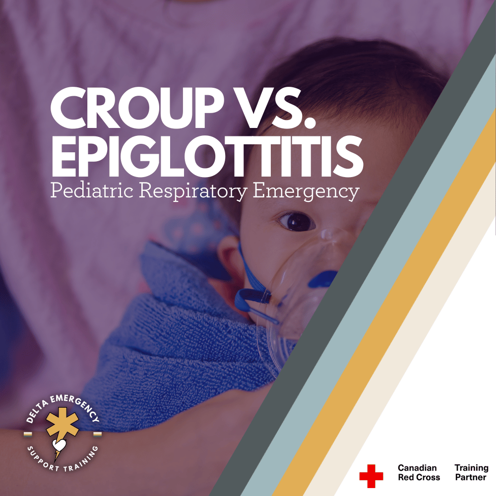
Croup vs. Epiglottitis in Children: What First Responders and Parents Need to Know

Breathing problems in children can be terrifying, and knowing the difference between croup and epiglottitis could save a life. Parents need to know the warning signs, while first responders must act quickly and carefully. Here’s how to tell them apart and what steps to take in an emergency.

When a child has trouble breathing, both parents and first responders feel the urgency. Two conditions often confused with one another are croup and epiglottitis. While both affect the airway, they differ dramatically in severity and required response. For parents, understanding the warning signs can help you know when to call for help. For first responders, recognizing the difference is critical to providing safe and effective care.

Croup: Common but Often Alarming

What it is:
Croup is a viral infection that causes swelling of the larynx and trachea, most often seen in children aged 6 months to 3 years.

Parents may notice:

  • Barking, seal-like cough (especially at night)

  • Hoarse voice

  • Stridor (high-pitched sound on inspiration) when crying or upset

  • Low-grade fever

  • Symptoms often start after a cold

First responder perspective:

  • Most cases are mild, but moderate to severe croup can cause stridor at rest, increased work of breathing (retractions, nasal flaring), or fatigue.

  • Treatment may include humidified oxygen, steroids (e.g., dexamethasone), and nebulized epinephrine in severe cases.

  • Keeping the child calm is key; agitation worsens obstruction.

Epiglottitis: A True Airway Emergency

What it is:
Epiglottitis is a bacterial infection that causes the epiglottis to swell rapidly, threatening complete airway obstruction. Thanks to Hib vaccination, it’s now rare, but still life-threatening when it occurs.

Parents may notice:

  • Sudden onset of high fever

  • Severe sore throat and difficulty swallowing

  • Drooling (child can’t swallow saliva)

  • Muffled voice

  • Child sitting forward, chin out, mouth open (“tripod position”)

  • Appearing very ill, anxious, and struggling to breathe

First responder perspective:

  • This is a do not delay, do not agitate situation.

  • Never attempt to look in the child’s throat — it can trigger complete obstruction.

  • Keep the child upright and calm.

  • Provide high-flow oxygen if tolerated.

  • Be prepared for advanced airway intervention (hospital setting or with ALS backup).

  • Rapid transport is essential.

Quick Comparison

FeatureCroup (Viral)Epiglottitis (Bacterial, Emergency)OnsetGradual, after cold symptomsSudden, rapid progressionCoughBarking, seal-likeRare or absentFeverLow-gradeHighVoiceHoarseMuffled, “hot potato” voiceSwallowingUsually ablePainful, droolingStridorCommon, louder when upsetLate, quieter, concerningSeverityUsually mild-moderateSevere, life-threatening

The Takeaway

For parents:

  • Croup is common and usually manageable, but if your child has stridor at rest, difficulty breathing, or bluish lips, call 911.

  • Epiglottitis is rare but an emergency — if you see drooling, high fever, tripod posture, or sudden severe breathing trouble, call for help immediately.

For first responders:

  • Stay calm and keep the child calm.

  • Differentiate based on history and presentation.

  • Supportive care is often enough for croup, while epiglottitis requires immediate rapid transport and careful airway management.

When it comes to pediatric airway conditions, the rule is simple: if in doubt, treat it like epiglottitis until proven otherwise. Quick recognition and action save lives.

Read More
Advanced First Aid, Awareness Kumari Malhotra Advanced First Aid, Awareness Kumari Malhotra

Distracting Injuries: Pulling Focus and Masking Pain

In trauma care, some injuries grab all the attention—but they aren’t always the deadliest. Distracting injuries can mislead responders and mask life-threatening conditions. Here’s what every first responder needs to know about spotting them, staying systematic, and keeping patient safety the top priority.

When trauma calls come in, it’s easy to fixate on the most obvious wound, the open fracture, the deep laceration, the heavy bleeding. But those are often distracting injuries: visible traumas that can obscure other, more dangerous threats. Recognizing them and continuing to assess systematically is what separates a good response from a lifesaving one.

What Is a Distracting Injury?

A distracting injury is one that draws your attention so strongly that both responders and patients may overlook other serious but less obvious issues. In paramedic protocols, these injuries can make a patient “unreliable” for clinical clearance of the cervical spine because they may mask neck pain or tenderness. As a result, spinal precautions should be maintained unless more definitive assessment rules them out.

Why They Matter

The danger of distracting injuries is twofold:

  1. They pull focus – A gruesome fracture or large bleed grabs everyone’s attention, often overshadowing a more subtle but life-threatening issue like a compromised airway or spinal injury.

  2. They mask pain – A patient in severe pain from a major injury may not report or even notice neck or back pain, leading responders to miss critical red flags for spinal trauma.

    Evidence-Based Insights

    Studies show distracting injuries are often upper-torso or limb injuries that change how a patient perceives or reports pain:

    • Heffernan et al. (2005) found that some patients with cervical spine fractures had no neck tenderness—but all had upper torso injuries like chest trauma, which likely distracted from the pain.

Common Examples

  • Long bone fractures (femur, humerus)

  • Major burns

  • Crushing or degloving wounds

  • Profuse bleeding or amputations

  • Severe chest or upper torso injuries

First Responder Priorities

  1. Stick to the primary survey (ABCs)
    No matter how distracting a wound looks, airway, breathing, and circulation must always come first.

  2. Protect the spine when in doubt
    If a distracting injury is present, assume the patient cannot reliably clear their spine. Apply spinal precautions unless imaging or protocols say otherwise.

  3. Reassess constantly
    A distracting injury may not only occupy the patient’s attention but yours as well. Don’t let tunnel vision take over.

  4. Think systematically, not emotionally
    The most dramatic injuries are not always the deadliest. Quiet but hidden injuries like internal bleeding or spinal cord compromise, may be far more dangerous.

The Bottom Line

Distracting injuries are more than just dramatic wounds—they can hide life-threatening problems and mislead both responders and patients. The best protection is discipline: follow a structured assessment, protect the spine when necessary, and never let your focus shift entirely to the injury that looks worst. A calm, systematic approach can make the difference between managing trauma and saving a life.

Read More

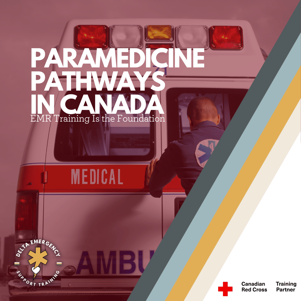
Becoming a Paramedic in Canada: How EMR Training Fits Into the Bigger Picture

Thinking about becoming a paramedic in Canada? It starts with understanding the paramedicine pathway. This blog explains where EMR training fits into the journey and why it’s a key first step toward a rewarding career in emergency services.

Across Canada, the demand for trained paramedics continues to rise. Communities rely on first responders not only for life-threatening emergencies but also for urgent care, transport, and support in times of crisis. If you’ve ever thought about a career in emergency services, you may be wondering: where do I start?

The answer for many is Emergency Medical Responder (EMR) training. This entry-level program provides the essential skills you need to help patients and also acts as a stepping stone into the professional paramedicine pathway. To understand why EMR is such a valuable first step, it helps to look at how the paramedicine system is structured in Canada.

1. The Paramedicine Ladder in Canada

In Canada, paramedicine is organized into different levels of training and certification. Each step builds on the one before it, adding more advanced knowledge, clinical skills, and responsibilities.

Emergency Medical Responder (EMR)

  • Entry-level certification recognized across many provinces.

  • Focuses on patient assessment, CPR and AED use, airway management, bleeding control, and trauma care.

  • EMRs may work in industrial safety, event medical services, rural response, and are often a requirement for admission into Primary Care Paramedic programs.

  • Training is usually short and focused, making it an accessible first step.

Primary Care Paramedic (PCP)

  • The first professional paramedic level in Canada.

  • PCPs are qualified to work on ambulances and provide advanced pre-hospital care.

  • Skills include medication administration, advanced airway management, and cardiac monitoring.

  • Typically requires 1–2 years of college-level education, depending on the province.

Advanced Care Paramedic (ACP)

  • Builds on the PCP scope of practice with advanced skills.

  • Includes procedures such as intubation, IV therapy, advanced cardiac interventions, and expanded medication options.

  • Training usually takes an additional 1–2 years beyond PCP.

Critical Care Paramedic (CCP)

  • The highest level of paramedic training in Canada.

  • Specializes in transporting and treating critically ill or injured patients, often by air ambulance or in hospital transfer situations.

  • CCPs provide intensive care support outside the hospital environment.

This tiered approach means that every paramedic, no matter how advanced, started with the same fundamentals—and that usually begins with EMR.

2. Why EMR Training Matters

Even though EMR is the entry-level program, it’s far more than a “basic” course. Here’s why it matters:

  • Foundational Skills: You’ll learn how to assess patients systematically, manage airways, perform CPR, use AEDs, and provide lifesaving first aid for medical and trauma emergencies.

  • Confidence in Emergencies: EMR training prepares you to step into real-world situations calmly and effectively, whether you’re responding at work, volunteering, or helping in your community.

  • A Career Sampler: Many people use EMR training as a way to “test drive” paramedicine before committing to the longer PCP or ACP programs. It’s a great way to discover if this career path is right for you.

  • Admission Requirement: In several provinces, completion of an EMR program is a mandatory prerequisite for applying to PCP programs.

In short, EMR is both a valuable certification on its own and a launching pad for more advanced paramedic training.

3. Career Opportunities After EMR

Some students complete EMR training because they want to work right away, while others use it as preparation for PCP school. Either way, opportunities exist.

  • Industrial and Remote Work: Oil and gas sites, mining operations, and construction companies often employ EMRs to provide on-site medical support.

  • Event Medical Services: Concerts, festivals, and sporting events need trained responders to manage emergencies on the ground.

  • Volunteer Roles: Many communities welcome EMRs as part of first responder programs, especially in rural or remote areas.

  • Pathway to PCP: EMR graduates have the foundation to move directly into Primary Care Paramedic education, making it a strategic step toward a long-term career.

4. The Canadian Context

Paramedicine is provincially regulated in Canada, which means that the scope of practice and educational requirements vary depending on where you live. For example:

  • In British Columbia and Alberta, EMR certification is a recognized entry point for working in certain EMS systems.

  • In Ontario, EMR may not be a licensed level, but training still provides an advantage for those entering PCP programs.

  • Across the country, EMR certification is widely respected as a sign of commitment to the field and readiness to pursue paramedic studies.

Regardless of the province, EMR training signals that you’ve mastered essential lifesaving skills and are prepared for the next step.

5. A Pathway, Not a Shortcut

One of the most important things to understand about paramedicine in Canada is that there are no shortcuts. Every level of training is rigorous and builds on the last. But EMR offers a pathway—an accessible starting point that opens doors, whether you want to work immediately or advance further.

By taking EMR, you’re not just learning a skillset. You’re joining a community of first responders who play a vital role in Canada’s healthcare system.

Conclusion

Becoming a paramedic in Canada is a journey. It starts with building a solid foundation—and for most students, that foundation is Emergency Medical Responder training. EMR equips you with lifesaving skills, gives you real-world confidence, and provides the stepping stone needed for higher levels of paramedicine like PCP, ACP, and CCP.

If you’re considering a career in emergency services, EMR training is the best way to begin. It’s the first step on a ladder of learning that leads to one of the most rewarding careers in healthcare.

👉 Ready to start your journey? Enroll in our EMR program today and take the first step toward becoming a paramedic in Canada.

Read More

ETCO₂: What It Is and Why It Matters for First Responders

When you first hear the term ETCO₂, it might sound like complicated medical jargon. But in reality, it’s a simple concept that every professional responder should understand — and once you do, it can completely change the way you see your patients.

Let’s break it down step by step.

What Does ETCO₂ Mean?

ETCO₂ stands for End-Tidal Carbon Dioxide.

  • End-Tidal = the very end of an exhaled breath.

  • Carbon Dioxide (CO₂) = the waste gas your body produces when it uses oxygen for energy.

So, ETCO₂ is literally the measurement of how much CO₂ is in the air a patient breathes out at the very end of their breath.

This number tells us an incredible amount about what’s going on inside the body — with both the lungs and the heart.

How Do We Measure ETCO₂?

ETCO₂ is measured using a device called capnography.

  • In simple terms, it’s a little sensor attached to a mask, nasal cannula, or an airway device.

  • It continuously analyzes the breath coming out and gives two things:

    1. A number (usually measured in mmHg, with normal being about 35–45 mmHg).

    2. A waveform (a little graph showing how the CO₂ rises and falls with each breath).

Why Is ETCO₂ Important?

Here’s the key: ETCO₂ reflects how well a patient is ventilating (moving air), but it also gives clues about circulationand metabolism. That’s why responders call it the “vital sign of ventilation.”

Think of it as a window into three systems at once:

  1. Airway & Breathing

    • Low or absent ETCO₂ can mean the patient isn’t breathing well, has an obstructed airway, or isn’t ventilated properly with a bag-valve mask.

  2. Circulation (Blood Flow)

    • In cardiac arrest, ETCO₂ is a powerful indicator of CPR quality. Good chest compressions circulate blood, and ETCO₂ rises.

    • A sudden spike in ETCO₂ can even mean return of spontaneous circulation (ROSC) — the patient’s heart has started beating again.

  3. Metabolism

    • Conditions like sepsis, diabetic emergencies, or shock can alter CO₂ levels. ETCO₂ helps responders piece together the bigger clinical picture.

Real-World Examples for Responders

  • Cardiac Arrest: ETCO₂ below 10 mmHg during CPR often means compressions aren’t effective. When it jumps above 35 suddenly, it may mean you’ve got ROSC.

  • Airway Management: If you intubate a patient and see a nice ETCO₂ waveform, you know the tube is in the trachea (not the stomach).

  • Respiratory Emergencies: In asthma or COPD, ETCO₂ waveforms can show “shark fin” patterns, helping you confirm and monitor the severity.

  • Sedation & Monitoring: If a patient is given pain medication, ETCO₂ helps detect if their breathing slows down before oxygen levels drop.

Why Should EMRs and Fire Applicants Care?

As an Emergency Medical Responder (EMR) or a firefighter applicant, understanding ETCO₂ gives you an edge. It shows you’re not just memorizing steps, but actually thinking about what’s happening inside the body.

  • It ties together your knowledge of the respiratory system and cardiovascular system.

  • It reinforces the importance of ventilation, circulation, and metabolic function.

  • And most importantly, it helps you make better decisions in high-pressure situations.

The Bottom Line

ETCO₂ might sound technical, but at its core it’s simple: it’s how we measure how well a patient is breathing and circulating. For responders, it’s one of the most valuable tools you can use to guide patient care, especially in emergencies where seconds matter.

At Delta Emergency Support Training, we break down concepts like ETCO₂ in plain language and then show you how to apply them in real-world scenarios. Our courses are taught by active paramedics and firefighters, so you’ll learn not just the “what,” but the “why” and “how” behind every skill.

Read More

Alberta Fire Applicants & EMRs: Master the Medical Portion with Paramedics and Firefighters

Succeeding in the medical portion of a firefighter or EMR application in Alberta comes down to mastering the fundamentals under pressure. At Delta Emergency Support Training, active paramedics and firefighters teach you vital patient assessment skills, accurate vitals, effective communication, and safe medication administration.

Why Fire Applicants Fail the Medical Portion (And How to Prepare)

Firefighting is one of Alberta’s most demanding careers, and the medical portion of the application ensures candidates can handle life-or-death situations both trauma and medical, under pressure. Many applicants struggle, not because they lack dedication, but because they underestimate the scenario-based, high-pressure nature of this part of the evaluation. Here’s what to know and how to prepare.

1. Not Understanding the Scenario-Based Format

Applicants may know first aid procedures but are unprepared for realistic simulations. Evaluators look for:

  • Correct prioritization of patient care

  • Efficient use of available equipment

  • Accurate assessment of vitals (pulse, blood pressure, respiration, oxygen saturation)

  • Maintaining composure under pressure

Preparation Tip:
Practice scenarios under timed conditions. Include taking accurate vitals for each patient, documenting them, and adjusting care accordingly. Repetition builds both speed and confidence.

2. Poor Time Management During Simulations

Time pressure is a common challenge during firefighter medical evaluations. Some applicants try to rush through tasks, either spending too long on minor details or speeding through essential steps. This can result in incomplete assessments, inaccurate vitals, missed medication checks, or poor patient communication.

Key Tip:

  • Don’t rush—take your time to work methodically.

  • Focus on the fundamentals of your patient assessment: scene safety → primary survey → airway, breathing, circulation → vitals → medications → secondary survey → ongoing care.

  • If you get stuck or feel pressured, return to the basics and systematically work through each step. Accuracy and thoroughness are more important than speed.

  • Use practice scenarios to balance efficiency with careful, precise assessment.

Preparation Tip:

  • Use a mental checklist: Scene safety → Primary survey → Secondary survey → Ongoing care → Hand-off report.

  • Practice completing your assessment efficiently while still accurately measuring vitals and administering medications.

  • Practice whenever you can! Run calls in your head.

3. Lack of Critical Thinking

Memorized procedures aren’t enough—patients don’t always behave as expected. Evaluators want to see adaptability: can you adjust your care based on vital signs, patient feedback, or medication needs?

Preparation Tip:

  • Train with scenarios including complications like fluctuating vitals, patient distress, or adverse medication reactions.

  • Ask yourself after each practice: “How would I adjust their oxygen saturations fall, or a medication has unexpected effects?”

4. Inadequate Patient Communication

Medical competence isn’t just technical—communicating clearly with patients is critical. Poor communication can make patients anxious or uncooperative, affecting assessment accuracy.

Preparation Tip:

  • Practice explaining what you’re doing and why while performing assessments and administering medications.

  • Use calm, clear language: “I’m going to give you your medication now; it may make you feel drowsy. Let me know if you feel unwell.”

  • Listen and respond to patient concerns—they may give vital clues about their condition.

5. Not Knowing Medications, Indications, and Contraindications

Many applicants fail by being unsure about common medications, their proper use, or when not to administer them. Evaluators want to see that you can safely give medications if required, understand dosage, and recognize contraindications.

Preparation Tip:

  • Study common pre-hospital medications: aspirin, epinephrine auto-injectors, nitroglycerin, oxygen, and glucose.

  • Know the indications (when to use), contraindications (when not to use), dosage, and potential side effects.

  • Practice scenario-based administration with simulated patients.

6. Inadequate Knowledge of Fire-Specific Medical Risks

Alberta applicants face unique hazards: industrial chemicals, wildfires, smoke inhalation, cold-weather trauma. Failing to anticipate these can affect performance.

Preparation Tip:

  • Focus on airway management, burns, smoke inhalation, rapid trauma assessment, vitals monitoring, and safe medication use.

  • Understand environmental impacts: extreme temperatures, long ambulance response times, and remote locations.

7. Overlooking Soft Skills and Professionalism

Evaluators notice panic, poor teamwork, and rushed or inaccurate communication. Both soft skills and technical skills are tested simultaneously.

Preparation Tip:

  • Role-play with peers, focusing on calm, professional communication.

  • Practice reporting vitals and medications verbally and accurately to simulate handoff to paramedics.

  • Debrief after every scenario: identify strengths and areas for improvement.

8. Tips for Final Preparation

  • Simulate Realistic Conditions: Wear PPE, gloves, and work under distractions to mimic real assessments.

  • Practice Accurate Vitals and Medication Use: Include pulse, respiration, oxygen saturation, blood pressure, and scenario-based administration of medications.

  • Time Your Scenarios: Track your completion time without sacrificing accuracy.

  • Review Fundamentals: CPR, airway management, bleeding control, shock recognition, spinal immobilization, and medications.

  • Debrief Every Practice: Discuss patient communication, vital trends, medication decisions, and overall decision-making.

Conclusion

The medical portion of the firefighter application isn’t meant to trip you up—it ensures you’re ready to handle emergencies. By practicing realistic scenarios, taking accurate vitals, communicating effectively with patients, knowing your medications, managing your time, and thinking critically, Alberta applicants can approach the evaluation confidently and maximize their chances of success.

At Delta Emergency Support Training, you’ll be taught by active paramedics and firefighters, gaining real-world insights and practical skills that go beyond the textbook. This hands-on instruction ensures you’re fully prepared for both the medical portion of your firefighter application and real-life emergencies.

Read More

Pulmonary Embolism for EMR Students: Signs, Symptoms, and Action Steps

Pulmonary embolisms are among the most serious complications post-surgery—and often go unnoticed until it’s too late. For Emergency Medical Responders, especially those working in Alberta’s high-risk environments, knowing the subtle signs and acting fast can make all the difference. This blog breaks down what to look for, how to respond, and why early recognition matters.

Pulmonary Embolism in Post-Surgical Patients

What Every Emergency Medical Responder Needs to Know

Pulmonary embolisms (PEs) are a leading cause of sudden, preventable death—especially in post-operative patients. As Emergency Medical Responders (EMRs) or Advanced First Aiders, understanding the signs, complications, and red flags of a PE can be the difference between life and death during a call.

While many PEs occur silently, those that do present can deteriorate rapidly. In the prehospital environment, your scene awareness, history taking, and rapid recognition play a crucial role in early intervention.

What Is a Pulmonary Embolism (PE)?

A pulmonary embolism is a blockage of one or more pulmonary arteries in the lungs, usually caused by a blood clot (thrombus) that travels from the legs (deep vein thrombosis or DVT) or pelvis. When that clot reaches the lungs, it obstructs blood flow, strains the heart, and impairs gas exchange—all of which can lead to hypoxia, shock, or cardiac arrest.

Why Are Post-Surgical Patients at Risk?

Surgical patients, particularly those recovering from major operations like orthopedic, abdominal, or pelvic procedures, face multiple risk factors:

  • Immobility during and after surgery → reduced circulation

  • Tissue trauma → activates the clotting cascade

  • Dehydration or blood loss → increases blood viscosity

  • Compression of veins during long procedures

  • Pre-existing conditions like obesity, smoking, cancer, or clotting disorders

Even young, otherwise healthy patients can develop PEs after surgery if proper precautions aren't followed.

Red Flags EMRs Should Watch For

A post-surgical patient complaining of vague symptoms may not look critical—but here are PE signs that demand immediate attention:

🫁 Respiratory Signs:

  • Sudden shortness of breath (unexplained)

  • Tachypnea (rapid breathing)

  • Chest pain (sharp, pleuritic — worsens with inspiration)

  • Cough (may be dry or produce blood-tinged sputum)

  • Low SpO₂ despite oxygen administration

❤️ Cardiovascular Signs:

  • Tachycardia

  • Hypotension

  • Lightheadedness or fainting

  • Cyanosis

  • Cool, clammy skin

🧠 Neurological & Other Signs:

  • Anxiety or impending sense of doom

  • Altered LOC in severe cases

  • Leg pain/swelling (DVT signs)

  • Recent history of surgery or long immobilization

Field Priorities for EMRs

  1. Scene Size-Up and History Taking

    • Ask about recent surgeries, travel, hospitalizations, or prior blood clots

    • Look for signs of immobility, compression devices, or anticoagulants

  2. Primary Assessment

    • Focus on airway, breathing, and circulation

    • Supplemental high-flow oxygen immediately if signs of hypoxia

  3. Vitals and Rapid Transport

    • Monitor vitals frequently; deterioration can be fast

    • Transport urgently with pre-alert to hospital if PE is suspected

  4. Reassessment

    • Watch for declining mental status or respiratory fatigue

    • Prepare for deterioration — cardiac arrest is possible in large PEs

What Makes PEs So Dangerous?

  • They can present subtly and escalate rapidly

  • Not all patients show textbook symptoms

  • Once the heart begins to strain or fail from the blockage, the window for intervention narrows quickly

  • Anticoagulant therapy or thrombolytics are hospital treatments, so early recognition and rapid transport are critical

Real-World Example

You’re dispatched to a 54-year-old woman 5 days post-knee replacement complaining of shortness of breath. She’s alert but anxious, SpO₂ is 88% on room air, heart rate 116, and she has mild leg swelling. She says it started suddenly and she feels “off.”

As an EMR, this is a classic red flag scenario: post-op, tachycardia, respiratory distress, possible DVT. This is when your training matters most.

Conclusion: What You Do Matters

Pulmonary embolisms in post-surgical patients are time-sensitive medical emergencies. As a responder, your ability to identify concerning symptoms, gather relevant history, and prioritize rapid care can drastically improve patient outcomes.

Always consider PE when a post-op patient presents with unexplained respiratory or circulatory compromise—even if it seems mild at first. Trust your instincts, act fast, and escalate early.

Delta Emergency Support Training

Based in Calgary, Alberta, we train EMRs and Advanced First Aiders with real-world scenarios and paramedic-led instruction to prepare you for critical cases like this. Whether you're working industrial, remote, or public events, our training prepares you for what truly matters.

Read More

Your Mental Checklist for Altered Level of Consciousness Calls

Altered level of consciousness (LOC) can signal everything from trauma to stroke, overdose, or diabetic emergencies. For EMRs and Advanced First Aiders, quick assessment using tools like AVPU and GCS is essential. Learn how to approach LOC calls with confidence and clinical clarity.

Altered level of consciousness (LOC) is a broad but critical presentation that can signal anything from head trauma to medical emergencies like hypoglycemia, stroke, or overdose. As an Emergency Medical Responder (EMR) or Advanced First Aider (AFA), your role in the initial assessment can directly influence the outcome for the patient.

These calls can be unpredictable, high-stress, and require fast decision-making. That’s why having structured tools like AVPU and GCS (Glasgow Coma Scale) in your mental toolbox is essential.

What Is Altered Level of Consciousness?

Altered LOC refers to any state where a person’s awareness, responsiveness, or orientation is impaired. It ranges from confusion or disorientation to full unresponsiveness.

Some common causes include:

  • Head trauma

  • Stroke or transient ischemic attack (TIA)

  • Seizures or postictal states

  • Hypoglycemia or hyperglycemia

  • Drug or alcohol overdose

  • Shock

  • Infection (e.g., sepsis or meningitis)

  • Psychiatric conditions

Because the causes are so diverse, your initial assessment plays a key role in identifying red flags, ruling out life threats, and communicating clearly with paramedics or dispatch.

Using the AVPU Scale

The AVPU scale is a quick and simple way to assess responsiveness in the field:

  • A – Alert: The person is awake and oriented to person, place, time, and event.

  • V – Verbal: The person responds to verbal stimuli but is not fully oriented.

  • P – Pain: The person does not respond to voice but reacts to pain (e.g., sternal rub).

  • U – Unresponsive: No response to voice or pain.

When to Use AVPU:

AVPU is ideal for initial assessments, especially during your primary survey. It gives you a quick snapshot of how serious the situation may be, helping prioritize transport and care.

Glasgow Coma Scale (GCS)

While AVPU is fast, the Glasgow Coma Scale provides a more detailed and objective assessment of neurological function.

The GCS evaluates three categories:

  1. Eye Opening (E) – out of 4

  2. Verbal Response (V) – out of 5

  3. Motor Response (M) – out of 6

CategoryScore BreakdownEye Opening4 – Spontaneous
3 – To voice
2 – To pain
1 – NoneVerbal Response5 – Oriented
4 – Confused
3 – Inappropriate words
2 – Incomprehensible sounds
1 – NoneMotor Response6 – Obeys commands
5 – Localizes pain
4 – Withdraws from pain
3 – Flexion (decorticate)
2 – Extension (decerebrate)
1 – None

GCS Scoring:

  • 13–15 = Mild impairment

  • 9–12 = Moderate

  • ≤8 = Severe; often indicates need for airway management

Even at the EMR or AFA level, familiarity with the GCS can help you provide accurate handovers and assist higher-level responders in decision-making.

Approach to the Call: LOC Management Steps

1. Scene Assessment & Safety

  • Scan for hazards (especially if drugs, trauma, or violent behavior suspected)

  • Note environmental clues (needles, alcohol, medications, crash helmets, etc.)

2. Primary Survey (ABCs)

  • Ensure Airway is open — unresponsive patients may need manual airway maneuvers or placement in recovery position.

  • Check Breathing — rate, effort, and quality.

  • Assess Circulation — skin signs, pulse, and potential bleeding.

3. Level of Consciousness Assessment

  • Use AVPU for rapid triage.

  • Apply GCS when appropriate and time allows.

  • Assess orientation to person, place, time, and event.

4. Vital Signs

  • Monitor pulse, respiration rate, blood pressure, temperature, SpO₂, and blood glucose (if within your scope).

5. Secondary Survey

  • Look for trauma (especially head/neck)

  • Check pupils (size, equality, reactivity)

  • Review SAMPLE history:

    • Signs and symptoms

    • Allergies

    • Medications (especially insulin, seizure meds, psych meds)

    • Past medical history

    • Last oral intake

    • Events leading up to incident

Communication Is Key

LOC calls often require rapid handover to EMS or transport to hospital. A strong report includes:

  • AVPU or GCS score

  • Vitals and changes in LOC

  • Pertinent SAMPLE history

  • On-scene findings (e.g., signs of overdose, trauma, seizure activity)

Using clear, clinical language shows professionalism and ensures continuity of care.

Why This Matters

Altered LOC may be the first sign of a life-threatening emergency. Whether you’re the first responder on a highway or treating someone at a workplace or remote site, your ability to assess, document, and respond effectively can help save a life—or prevent rapid deterioration.

Train with Confidence at Delta Emergency Support Training

At Delta Emergency Support Training in Calgary, our Advanced First Aid and EMR-level courses prepare students to manage complex scenes—including those involving altered LOC. Taught by experienced paramedics and firefighters, our training focuses on real-world decision-making, hands-on assessments, and scenario-based practice.

Whether you're renewing your skills or building toward a career in EMS or firefighting, we’re here to help you respond with clarity, confidence, and care.

Read More
Alberta, Advanced First Aid, Certificates Kumari Malhotra Alberta, Advanced First Aid, Certificates Kumari Malhotra

From Oilfield to Firehall: Why Tradespeople Are Switching to Emergency Services

Many tradespeople in Alberta’s oil and gas sector are transitioning to emergency services careers. At Delta Emergency Support Training in Calgary, our Advanced First Aid course provides the essential skills and certification needed to make that move with confidence.

Alberta has long been known for its oil and gas industry — and rightly so. From Fort McMurray to the foothills outside Calgary, thousands of workers have built careers in the trades, on the rigs, and in the field. But in recent years, a growing number of tradespeople are making a dramatic shift: trading in steel toes and coveralls for bunker gear and medical kits.

At Delta Emergency Support Training, based in Calgary, Alberta, we’ve seen the trend firsthand. More and more of our Advanced First Aid (AFA) students come from backgrounds in the oil patch, construction, or related trades, seeking a new path in firefighting, emergency medical services, or public safety.

Here’s why this transition is becoming so popular—and how Advanced First Aid training can be your first step from the oilfield to the firehall.

Why Tradespeople Are a Natural Fit for Emergency Services

Many of the qualities that make someone successful on a rig or in a trade are the same qualities needed in emergency response:

  • Strong work ethic and long shifts

  • Team-based environments

  • High-pressure problem solving

  • Hands-on technical skills

  • Risk awareness and safety training

  • Comfort in unpredictable or hazardous settings

If you’ve spent time working in the field, you’ve likely already experienced critical thinking during crises, emergency evacuations, or injury responses. That mindset—and real-world toughness—is highly transferable to fire and EMS careers.

Why the Shift Is Happening Now

There’s no doubt the oil and gas industry still offers high-paying work. But for many tradespeople, the physical demands, economic uncertainty, and cyclical layoffs have made them look for more stable, meaningful, or community-focused careers. Emergency services are standing out for a number of reasons:

  • Long-term stability and benefits

  • Purpose-driven work that gives back to the community

  • Opportunity to work closer to home

  • Union support and career advancement pathways

  • Strong demand for trained emergency responders in Alberta

If you're already safety-certified, used to remote work, and calm under pressure, transitioning to a fire department or EMS service might feel like a natural progression.

Advanced First Aid: Your First Step Toward a New Career

At Delta Emergency Support Training, we offer Advanced First Aid certification and recertification, designed for those ready to pursue new roles in emergency services.

Our courses, taught by experienced paramedics and firefighters, give you the skills and credentials to apply to:

  • Municipal fire departments like Calgary or Edmonton Fire

  • Industrial or wildland fire crews

  • Emergency Medical Responder (EMR) or Primary Care Paramedic (PCP) programs

  • Oil and gas site medical teams

  • Search and Rescue or auxiliary fire departments

What You’ll Learn:

Following the Canadian Red Cross curriculum, Advanced First Aid training includes:

  • Scene and patient assessment

  • CPR Level C and AED

  • Airway and breathing emergencies

  • Spinal, head, chest, abdominal, and limb injuries

  • Seizures, strokes, anaphylaxis, diabetic emergencies

  • Multiple casualty management and triage

  • Oxygen administration

  • Legal responsibilities and assisting with medications

This level of training is often the minimum requirement for entry into emergency service programs—and it builds confidence for high-stakes environments.

You're Not Starting Over—You're Building Forward

Switching careers doesn’t mean leaving your skills behind. In fact, your experience in the trades—whether it's welding, electrical, drilling, construction, or industrial safety—equips you with a unique edge.

At Delta Emergency, we’ve seen all kinds of professionals make the leap into emergency services. We’ve trained doctors, nurses, personal trainers, military personnel, and tradespeople who have successfully transitioned into fire, EMS, or rescue careers. The one thing they all had in common? A willingness to start with solid foundational training and a drive to serve.

You don’t need a medical degree or fire academy certificate to get started—just the right training and mindset.

Take the First Step with Delta Emergency Support Training

Emergency services are more than just a job—they're a calling. If you're ready to explore a career that's local, stable, and community-centered, Delta Emergency Support Training in Calgary, Alberta is here to support your journey.

Advanced First Aid is more than a certificate—it’s your starting point. Learn from real paramedics and firefighters and take the first step toward a career with purpose.

Visit our website to learn more about the program and how it can support your next move.

Read More

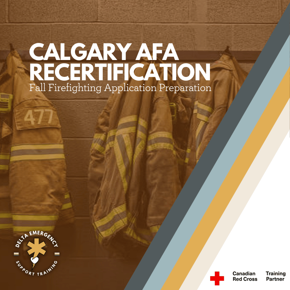
Planning to Apply to Edmonton or Calgary Fire? Why Advanced First Aid Recertification Matters

Planning to apply to Calgary or Edmonton Fire? Delta Emergency Support Training in Calgary offers Advanced First Aid recertification taught by paramedics and firefighters. Choose from weekday or weekend formats between September and November. Build your resume and keep your skills current with this Red Cross-certified course.

If you’re preparing to apply to Calgary or Edmonton Fire Department, you already know how competitive the process can be. These fire services are among the most respected in the country, and applicants are expected to show up well-prepared—both physically and professionally. One key credential that helps you stand out? Advanced First Aid (AFA) certification.

At Delta Emergency Support Training, located in Calgary, Alberta, we offer Advanced First Aid recertification courses taught by experienced paramedics and firefighters. Whether you’re updating your qualifications or preparing for pre-employment testing, our Red Cross-certified courses are designed to build the skills today’s fire services expect.

Why Advanced First Aid Is So Important

Advanced First Aid is not just a requirement—it’s a critical part of the emergency toolkit for any aspiring firefighter. It equips you to respond effectively to medical emergencies in both isolated and urban environments.

Departments like Calgary and Edmonton Fire often look for applicants who:

  • Hold a current Red Cross Advanced First Aid certification

  • Can demonstrate hands-on medical response experience

  • Have up-to-date knowledge of patient care, trauma management, and scene safety

Even if you've already taken the course before, recertifying ensures your skills meet current standards. Red Cross AFA certificates are valid for three years, but many departments recommend recertifying every two years to stay sharp and competitive.

What You’ll Learn in the Red Cross AFA Recertification Course

Our Advanced First Aid recertification course follows the Canadian Red Cross curriculum, providing practical training rooted in the latest national guidelines. Course content includes:

  • Scene assessment and patient surveys (primary and secondary)

  • Airway management and oxygen therapy

  • CPR Level C and AED usage

  • Treatment of traumatic injuries: spinal, chest, head, abdominal, and limb injuries

  • Medical emergencies: diabetic crises, seizures, allergic reactions, strokes, and more

  • Environmental emergencies: heat-related illnesses, cold exposure, poisonings

  • Multiple casualty management and triage

  • Legal considerations and documentation

  • Assisting with emergency medications

This course is designed for individuals who need advanced-level first aid for employment, volunteer service, or career advancement in emergency services.

Course Formats That Fit Your Schedule

We understand that many of our students are working professionals or balancing training, volunteering, and job applications. That’s why we offer two course delivery options:

Weekday Intensive Option

  • Monday to Friday

  • 09:00 to 18:00

  • Complete your recertification in just one week

Weekend Format

  • Friday evenings 18:00 to 22:00

  • Saturdays and Sundays 09:00 to 18:00

  • Spread across multiple weekends

Both formats offer full Red Cross certification and the same high-quality instruction led by frontline emergency responders.

Course Dates: September to November

We offer recurring Advanced First Aid recertification courses from September through November. All upcoming course dates are posted on our registration page. Early registration is recommended as seats fill quickly—especially ahead of common firefighter application and testing periods.

Build Your Resume While You Prepare

Recertifying in Advanced First Aid is one of many ways to strengthen your application. Other ways to build your fire service resume include:

  • Volunteering with local Search and Rescue, Fire Cadet, or community safety programs

  • Completing relevant certifications like NFPA 1001, ICS-100, Wildland Firefighter Training, or CPR Instructor

  • Gaining experience in related jobs such as lifeguarding, EMS, security, or industrial safety

  • Preparing for CPAT or A-PREP testing

Ready to Take the Next Step?

Whether you’re applying this year or planning ahead, Delta Emergency Support Training in Calgary is here to help you meet your goals. Our Advanced First Aid recertification courses are built to help aspiring firefighters like you gain the knowledge, confidence, and credentials you need to succeed.

Spots are limited and fill quickly as fire recruitment ramps up each fall.

Read More