The Delta Dispatch
The Lymphatic System: Your Body’s Built-In Defense Network
When most people think about vital systems in the human body, they picture the heart pumping blood or the lungs drawing in oxygen. But behind the scenes, the lymphatic system plays a critical and often overlooked role in keeping us healthy—especially when it comes to fighting infection, maintaining fluid balance, and supporting the immune system.
For first aiders, EMRs, and other frontline responders, understanding the lymphatic system can deepen your knowledge of the body's response to injury, infection, and inflammation. It also helps explain why we sometimes see swollen lymph nodes, fevers, and inflammatory reactions during patient assessments.
🔬 What Is the Lymphatic System?
The lymphatic system is a complex network of vessels, nodes, organs, and tissues that work together to:
Drain excess fluid from tissues and return it to the bloodstream
Filter harmful substances, like bacteria, viruses, and toxins
Produce and transport immune cells, such as lymphocytes
Absorb and transport fats from the digestive system
Think of it as the body’s sanitation and security team, quietly working alongside the circulatory system.
🧠 Key Components of the Lymphatic System
1. Lymph Fluid
A clear or pale yellow fluid made up of water, proteins, white blood cells (especially lymphocytes), and waste products.
Forms from fluid that leaks out of capillaries into tissues (called interstitial fluid) and is then collected by lymphatic vessels.
2. Lymphatic Vessels
A network of thin tubes that run throughout the body, similar to veins.
These vessels carry lymph fluid in one direction—toward the heart—using valves and muscular contractions to keep the fluid moving.
They eventually drain into two major ducts:
Thoracic duct (left side of body)
Right lymphatic duct (right side of head, arm, chest)
3. Lymph Nodes
Small, bean-shaped structures located along lymph vessels.
Filter lymph fluid and trap pathogens, foreign particles, and cancer cells.
Contain immune cells (B cells and T cells) that destroy harmful substances.
Common clusters are found in the neck, armpits, and groin—these may swell during infections.
4. Lymphoid Organs
Spleen: Filters blood, removes old red blood cells, and stores white blood cells.
Thymus: Located in the chest; where T cells mature (especially active in children).
Tonsils and adenoids: Located in the throat; protect against pathogens entering via mouth and nose.
Peyer’s patches: Found in the small intestine; monitor intestinal bacteria and immune responses in the gut.
🛡️ Lymphatic System and Immunity
The lymphatic system is deeply integrated into the immune response. It helps the body recognize, attack, and eliminate threats like viruses, bacteria, and cancer cells.
Key immune system cells found in lymphatic tissues include:
B lymphocytes (B cells): Produce antibodies to neutralize pathogens.
T lymphocytes (T cells): Attack infected cells and coordinate immune responses.
Macrophages: Engulf and digest cellular debris, pathogens, and dead cells.
When the body detects an invader, immune cells gather in nearby lymph nodes—causing localized swelling and tenderness, which you might feel in the neck or armpit during illness.
💉 Lymphatic System in Emergency Medicine and First Aid
While the lymphatic system itself doesn’t often present as a primary emergency, it plays a supporting role in many situations:
1. Infection
Swollen, tender lymph nodes can signal localized or systemic infection.
This is common in throat infections, skin abscesses, or infected wounds.
2. Sepsis
When infection spreads into the bloodstream, the lymphatic system may become overwhelmed.
Understanding its role helps responders recognize systemic responses like fever, elevated heart rate, and altered mental status.
3. Cancer
Lymph nodes are often used in staging cancers, as some malignancies (e.g., lymphoma or breast cancer) spread via the lymphatic system.
Responders may encounter patients with known lymphatic involvement or lymphedema from past cancer treatment.
4. Trauma and Swelling
Impaired lymph drainage can contribute to edema in injured limbs.
Compression injuries, surgical removal of nodes, or severe inflammation may damage lymphatic flow.
🧠 Fast Facts for First Aiders and EMRs
The lymphatic system has no central pump like the heart; it relies on movement, breathing, and muscle contractions.
Swollen lymph nodes are usually not painful unless infected.
Lymph nodes larger than 1 cm that persist for more than a few weeks should be evaluated by a physician.
Lymph fluid eventually returns to the bloodstream, helping maintain fluid balance and blood volume.
Conditions like lymphedema, often seen in cancer survivors, are caused by damage or blockage in lymph vessels.
🩺 Support the System
While you won’t be treating the lymphatic system directly in most emergency calls, here are ways first responders support it indirectly:
Encourage early treatment of infections to reduce the burden on lymph nodes
Recognize swollen lymph nodes as a sign of localized infection
Position patients properly to promote lymph drainage and circulation
Understand underlying conditions like lymphoma or lymphedema when assessing patients
📚 Conclusion: Small System, Big Impact
The lymphatic system may be quiet, but it’s crucial—working around the clock to remove waste, protect against illness, and support your immune defenses. For first responders, a working knowledge of the lymphatic system helps sharpen your assessment skills, especially in cases involving fever, infection, or unexplained swelling.
Whether you’re taking Advanced First Aid or working as an EMR, understanding this system adds another layer to your ability to assess and care for patients holistically.
Advanced First Aid for Dog Bites: A Firefighter & EMS Responder’s Guide
Dog bites demand swift, skilled care from first responders. Our Red Cross Advanced First Aid & EMR course trains firefighters and EMS personnel in critical skills—from scene safety to wound management and emotional support. Learn to confidently handle dog bite emergencies and more with hands-on training that prepares you for real-world situations.
Dog bites are common and can range from minor injuries to life-threatening wounds. As an advanced first aider—often a firefighter or EMS responder—you’re frequently the first healthcare professional on scene. Your response can significantly impact the victim’s recovery, infection risk, and emotional well-being.
This guide provides an in-depth approach to managing dog bites, emphasizing scene safety, injury treatment, wound care, and patient support with your advanced training and EMS protocols in mind.
1. Scene Safety: Your First Priority
As firefighters and EMS responders, scene safety is non-negotiable. Before attending to the victim:
Ensure the dog is secured or removed from the scene.
Coordinate with police or animal control if the dog is aggressive or unrestrained.
Use situational awareness to spot hazards like traffic, bystanders, or multiple animals.
Wear appropriate personal protective equipment (PPE), including gloves and eye protection.
Brief your team on potential risks and establish safe zones.
Remember: your safety is critical. You cannot assist others if injured.
2. Rapid Assessment of Injury and Vital Signs
Upon approaching the patient:
Perform a quick but thorough assessment:
Identify bite locations, wound depth, and bleeding severity.
Assess airway, breathing, circulation (ABCs), especially if bites are on the face or neck.
Look for signs of shock or distress.
Evaluate distal circulation and neurological status if extremities are involved.
Prepare for rapid transport if injury severity or patient condition warrants.
3. Managing Bleeding and Stabilizing the Wound
Apply direct pressure with sterile dressings to control bleeding.
Elevate the injured limb if it does not exacerbate pain or injury.
Use hemostatic dressings or pressure bandages if standard methods fail and bleeding is life-threatening.
Avoid closing wounds—leave that to hospital staff.
If bones are exposed or fractures suspected, immobilize the limb appropriately.
4. Cleaning and Infection Control in the Field
Use available clean water or saline to gently irrigate wounds, flushing away debris and contaminants.
Avoid harsh scrubbing to prevent tissue damage.
Apply sterile, non-adherent dressings loosely to reduce infection risk.
Maintain standard precautions—dog bites pose high infection risk from mixed bacterial flora.
5. Addressing Emotional Trauma and Providing Reassurance
Understand that dog bite victims, especially children, often experience intense fear or shock.
Use your training in crisis intervention to calm and reassure patients and caregivers.
Maintain eye contact, speak in a calm voice, and explain procedures as you go.
If a child is involved, engage a parent or guardian for comfort, and use age-appropriate communication.
6. Differences in Treatment: Adults vs. Children
Children’s smaller anatomy and thinner skin increase risk of deeper injuries. Facial and neck bites require immediate airway assessment.
Children may be less cooperative; use gentle distraction and clear, comforting language.
Monitor for airway obstruction and signs of respiratory distress in both adults and children, but be especially vigilant with pediatric patients.
Adults may underreport pain or injury severity—conduct thorough assessments.
7. When to Activate Advanced Care and Transport
Immediately initiate rapid transport and notify receiving facility if:
Bleeding is uncontrolled or severe
Patient shows signs of shock or compromised airway
Wounds involve face, neck, hands, or joints
Suspected fractures or nerve injuries are present
Signs of infection or systemic illness are evident
Pediatric patients require specialized care
Coordinate transport with EMS and hospital staff for optimal continuity of care.
8. Documentation and Reporting
Record detailed information on the bite incident, dog description, and circumstances.
Document patient’s vital signs, treatment provided, and response to interventions.
Share info with receiving medical personnel and law enforcement/animal control as needed.
Proper documentation supports patient care and public safety.
9. Post-Incident Patient Education
Advise victims to seek follow-up care for tetanus boosters, rabies prophylaxis, and wound management.
Educate on signs of infection to watch for at home.
Stress importance of reporting dog bites to local health authorities.
10. Summary for Firefighters & EMS Responders
StepKey ActionsScene SafetySecure environment, coordinate with animal control, PPE use.AssessmentRapid injury and ABC evaluation, assess for shock.Bleeding ControlDirect pressure, elevation, hemostatic dressings if needed.Wound CareGentle irrigation, sterile dressing, infection prevention.Emotional SupportCalm communication, involve caregivers, monitor for shock.Pediatric CareAirway vigilance, gentle approach, watch for distress signs.TransportRapid evacuation for severe cases, notify hospital.DocumentationAccurate records for care and reporting.
As a firefighter or EMS advanced first aider, your training equips you to manage the complex needs of dog bite victims with confidence and professionalism. Your timely intervention saves lives, prevents complications, and supports recovery—physically and emotionally.
How to Manage Delirium in Geriatric Patients: Essential Skills for EMRs and Firefighters
Delirium is an acute medical condition that often affects geriatric patients, causing confusion, disorganized thinking, and altered levels of awareness. As an EMR or firefighter, it's crucial to identify delirium early and manage it effectively. This guide explains how to assess, treat, and respond to elderly patients showing signs of delirium, ensuring optimal care in critical situations.
Delirium in the Geriatric Patient: How EMRs and Firefighters Can Identify and Respond
Delirium is a serious medical condition, particularly in the elderly, and as an EMR or firefighter, it’s crucial to recognize the signs early and act swiftly. Unlike dementia, which progresses slowly over time, delirium can develop acutely and rapidly change the patient’s mental status, presenting significant challenges for first responders. In geriatric patients, delirium is a medical emergency that often requires urgent intervention.
This blog will guide you on how to recognize delirium, understand its underlying causes, and effectively manage geriatric patients experiencing it.
What is Delirium?
Delirium is defined as an acute and fluctuating onset of inattention, disorganized thinking, and/or altered levels of awareness. It differs from dementia in that it is temporary and occurs suddenly, often in response to an underlying medical condition or environmental trigger.
The symptoms of delirium typically appear suddenly and may fluctuate throughout the day. Common signs include:
Inattention: Difficulty maintaining focus, distracted, or unable to follow a conversation.
Disorganized Thinking: Incoherent or illogical speech, trouble forming coherent thoughts.
Altered Awareness: The patient may be either overly drowsy (hypoactive) or agitated (hyperactive). In many elderly patients, hypoactive delirium is most common and can often go undetected in emergency situations.
Why is Delirium Common in Geriatric Patients?
The elderly population is particularly susceptible to delirium for several reasons, including:
Age-Related Physical Changes: Aging can cause changes in the brain that make older adults more vulnerable to confusion and mental disturbances.
Underlying Medical Conditions: Chronic health issues like heart disease, diabetes, or kidney failure may predispose the elderly to developing delirium.
Medications: Certain medications, especially in combination, can trigger delirium in older patients. Common culprits include sedatives, narcotics, and anticholinergics.
Infections: Conditions like urinary tract infections (UTIs) and pneumonia are known triggers of delirium in the elderly.
Environmental Factors: Hospitalizations, changes in routine, or unfamiliar environments (e.g., new settings after an injury) can exacerbate confusion and lead to delirium.
Recognizing Delirium in the Elderly
As an EMR or firefighter, recognizing delirium is a crucial skill that can improve patient outcomes. Delirium may not be as obvious as other acute conditions, especially since the elderly are often at risk of hypoactive delirium, which can manifest as lethargy or withdrawal rather than agitation.
Key Symptoms to Look For:
Inability to focus: The patient may appear "spacey," unable to follow a conversation, or may forget what they were just talking about.
Confusion: They may be disoriented, unable to recall where they are, or confused about the time and date.
Fluctuating alertness: The patient's level of awareness may change rapidly, ranging from excessive sleepiness to periods of hyperactivity or aggression.
Hallucinations or Delusions: While less common, some patients may see or hear things that aren’t there, or they may have false beliefs (e.g., believing they’re in a completely different location).
Hypoactive Delirium: A Hidden Danger
It’s important to note that 70% of delirium cases in elderly patients are hypoactive, meaning the patient may seem unusually drowsy, withdrawn, or lethargic. This subtle form can often go undiagnosed in the emergency department (ED) or in the field because the patient may not exhibit the typical signs of agitation. As a first responder, you must be aware that any older adult exhibiting lethargy or confusion after a recent trauma or medical event could be suffering from delirium.
Common Causes of Delirium in the Geriatric Patient
Several medical conditions or factors may trigger delirium in older adults, making them more vulnerable to changes in their cognitive state. As an EMR or firefighter, understanding these causes can help you quickly identify potential underlying issues:
Infections: UTIs, pneumonia, or sepsis can quickly lead to delirium in elderly patients.
Dehydration and Malnutrition: Inadequate fluid or nutrition intake can significantly affect brain function.
Medications: Polypharmacy, or the use of multiple medications, is common in elderly patients and can interact to cause confusion or delirium. Pay special attention to narcotics, sedatives, and antihistamines.
Pain or Injury: Trauma, especially head injuries, can trigger delirium, as can untreated pain.
Hospitalization: Elderly patients who are hospitalized for an acute illness or injury may experience delirium due to the stress of hospitalization, changes in routine, and medication side effects.
Alcohol Withdrawal: If the elderly patient has a history of alcohol use and suddenly stops drinking, delirium may occur.
How EMRs and Firefighters Can Respond to Delirium in the Elderly
When faced with a patient exhibiting signs of delirium, quick and effective action is critical. Below are steps for managing these patients:
1. Initial Assessment
Ensure Safety: The first priority is ensuring the safety of the patient. Patients experiencing delirium may be disoriented or agitated, so they could put themselves or others at risk. Ensure a safe environment and avoid sudden movements that might escalate confusion.
Take a Comprehensive History: If possible, gather a thorough history from caregivers, family, or bystanders. Ask about the patient’s recent health changes, medications, and any prior cognitive issues.
Rule Out Life-Threatening Causes: Delirium can be a sign of a serious condition such as an infection, stroke, or head injury. Check vital signs and assess for any signs of a critical underlying cause (e.g., hypotension, fever, abnormal heart rate, etc.).
2. Address Underlying Causes
If the delirium is due to a known cause (e.g., an infection), it is critical to treat the underlying issue as soon as possible.
Consider the possibility of hypoxia, hypoglycemia, or medication-related causes, which can often be quickly addressed in the field.
If the patient is in pain or has a known medical condition that could be contributing to confusion (like dehydration), take steps to manage it immediately.
3. Monitor the Patient
If the patient is not yet stable, keep a close watch on their mental status. Delirium can fluctuate, so continuous assessment is key.
Stay calm and provide reassurance to the patient. If they are agitated, offer clear explanations and avoid confrontational responses.
Transport to the hospital promptly, ensuring that the medical team is aware of the patient’s delirium so they can investigate and treat the underlying cause quickly.
4. Engage Family or Caregivers
When possible, engage family members or caregivers to help assess the patient's baseline mental status and to provide relevant medical history, including recent changes or new medications.
Conclusion
Delirium in geriatric patients is a common, often undiagnosed condition that presents a unique challenge for EMRs and firefighters. Because it can rapidly change from one moment to the next, it's essential to recognize the signs of delirium early and manage the underlying causes. Prompt action, effective communication with medical teams, and ensuring patient safety are key components in improving patient outcomes.
As a first responder, your ability to identify delirium and intervene early can mean the difference between a successful recovery and long-term complications. By staying vigilant and informed, you can ensure that older patients receive the timely, life-saving care they need.
Delta Emergency is taught by a well-versed group of instructors and owners who are experienced paramedics. We teach you not only the necessary skills but also how to be prepared for real-life emergencies. Our hands-on training ensures you're equipped to handle any critical situation with confidence, improving both your ability to respond and your patient outcomes.
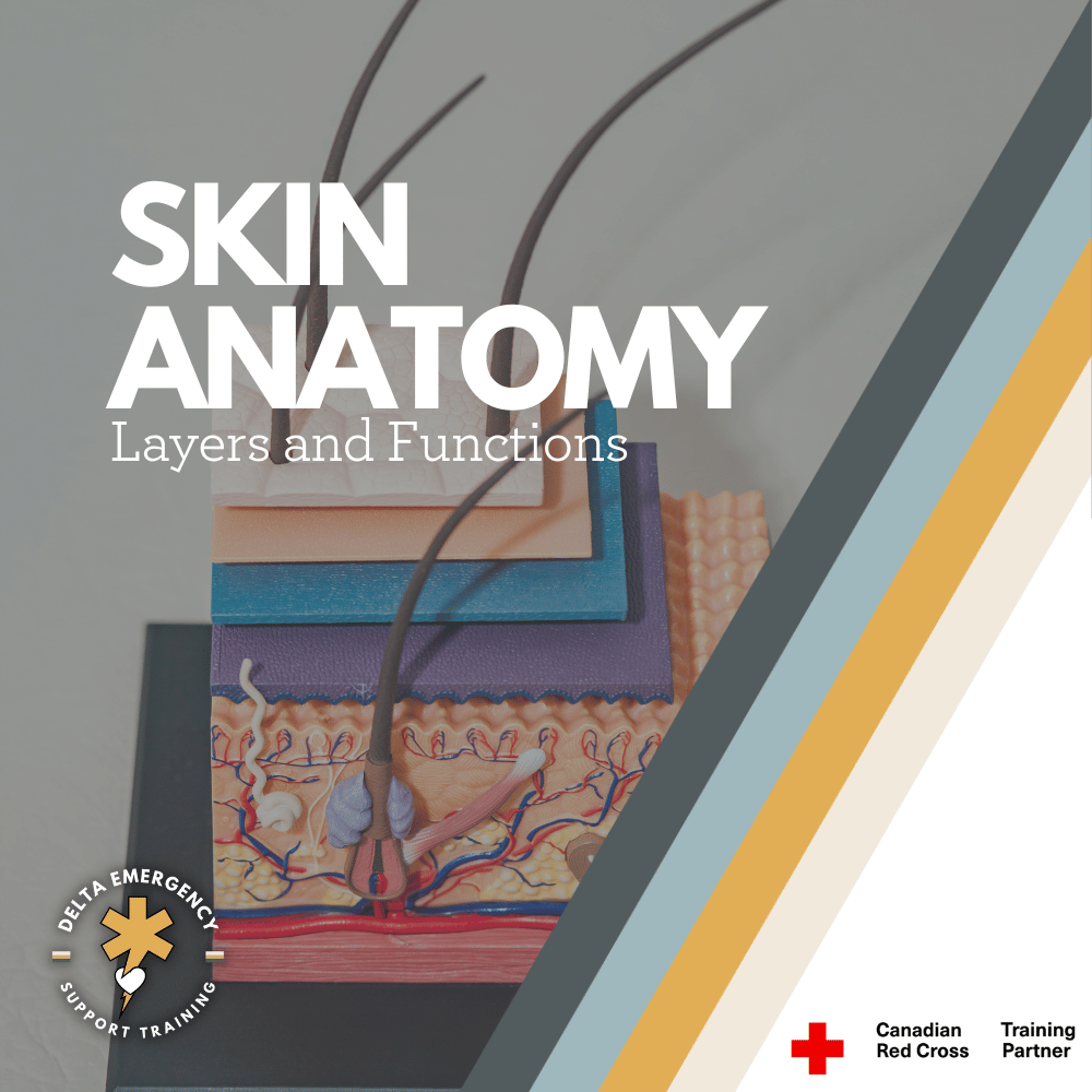
Skin Anatomy Overview for First Responders: Layers and Functions
Understanding the anatomy of the skin is essential for first responders. The skin serves as the body’s first line of defense, regulating temperature and protecting against infection. In this blog, we’ll break down the three layers of skin—epidermis, dermis, and hypodermis—and explore their functions, common injuries, and how this knowledge can help you provide better care in emergency situations.
As a first responder, you're trained to respond to a variety of emergencies, from burns and cuts to more complex trauma situations. Understanding the anatomy of the skin is crucial, as it plays a vital role in protecting the body from infection, regulating temperature, and healing injuries. In this blog, we’ll break down the layers of the skin, their functions, and how this knowledge helps you in your critical role as a first responder.
medical skin layer model
The Basics of Skin Anatomy
The skin is the body's largest organ, serving as the primary barrier between the internal systems and the outside world. It protects against physical damage, pathogens, and dehydration, while also playing a key role in temperature regulation. The skin is made up of three main layers, each with its own distinct function:
Epidermis (Outer Layer)
Dermis (Middle Layer)
Hypodermis (Subcutaneous Layer)
1. The Epidermis: The Body’s First Line of Defense
The epidermis is the outermost layer of skin, providing a waterproof barrier that shields the body from harmful substances, bacteria, and UV radiation. As a first responder, you will often encounter patients with injuries to the epidermis, such as cuts, abrasions, burns, or rashes.
Functions of the Epidermis:
Protection: The epidermis acts as the first line of defense against pathogens, chemicals, and physical damage.
Barrier to Water Loss: It helps prevent excessive loss of moisture, maintaining hydration in the body.
Skin Pigmentation: Melanin, produced in the epidermis, helps protect deeper layers from UV damage.
Sensation: The epidermis contains nerve endings that allow the skin to respond to environmental stimuli, like heat or pressure.
Important Considerations for First Responders:
Burns: Since the epidermis contains no blood vessels, burns typically affect this layer first, causing redness, swelling, and pain. In severe cases, it may also affect the dermis, leading to more serious injuries.
Infections: When the epidermis is compromised (such as in open wounds), there’s a higher risk of infection. Proper cleaning and dressing of wounds are essential to avoid bacterial infiltration.
2. The Dermis: The Skin’s Strength and Support
The dermis lies beneath the epidermis and is responsible for most of the skin's strength, elasticity, and sensation. This layer contains blood vessels, nerves, hair follicles, sweat glands, and connective tissue.
Functions of the Dermis:
Structural Support: The dermis contains collagen and elastin, proteins that provide strength and elasticity to the skin, allowing it to stretch and return to its original shape.
Temperature Regulation: Blood vessels in the dermis help regulate body temperature by constricting to conserve heat or dilating to release heat.
Sensation: The dermis is rich in nerve endings that detect pain, pressure, temperature, and touch.
Wound Healing: The dermis plays a critical role in healing skin wounds, as it contains the blood vessels and connective tissue that support tissue repair.
Important Considerations for First Responders:
Deep Cuts and Lacerations: When treating deep cuts or lacerations, you may encounter injury to the dermis, which could result in more severe bleeding due to the presence of blood vessels.
Blisters and Burns: Severe friction or thermal injuries can cause fluid buildup between the epidermis and dermis, leading to blisters. Burns can also extend into the dermis, causing damage to blood vessels and deeper tissues.
3. The Hypodermis: The Body’s Cushion and Insulator
The hypodermis, or subcutaneous layer, lies below the dermis and is primarily made of fat and connective tissue. This layer plays a significant role in protecting internal organs, insulating the body, and storing energy.
Functions of the Hypodermis:
Insulation: The fatty tissue helps regulate body temperature by providing insulation against heat loss.
Cushioning: It acts as a shock absorber, protecting bones and organs from injury by absorbing impact.
Energy Storage: The hypodermis stores fat, which serves as an energy reserve for the body.
Anchoring the Skin: It helps anchor the skin to underlying muscles and bones, giving the skin its flexibility and strength.
Important Considerations for First Responders:
Severe Trauma: In cases of deep trauma, such as blunt force injuries, the hypodermis may be damaged. This could lead to bruising or deeper tissue damage, as blood vessels in the hypodermis may rupture.
Frostbite: The hypodermis is the most affected area during frostbite, as it is the deepest layer. Severe cold temperatures can cause the fat cells to freeze, potentially leading to tissue death and the need for immediate medical intervention.
Skin Injuries and First Aid: Key Points for First Responders
As a first responder, understanding the anatomy of the skin helps you assess injuries more effectively and provide better care. Here are some common skin injuries and how they relate to the skin’s structure:
Abrasions: These involve damage to the epidermis, often caused by friction. While they are generally not severe, they can lead to infection if not properly cleaned and dressed.
Lacerations: Deep cuts can damage both the epidermis and dermis, leading to bleeding and pain. Proper wound care includes stopping the bleeding, cleaning the wound, and applying appropriate dressings.
Burns: Burns can range from superficial (affecting only the epidermis) to severe (penetrating the dermis and hypodermis). First-degree burns may cause redness and pain, while second-degree burns result in blisters and more intense pain. Third-degree burns involve damage to all layers of the skin and require immediate medical attention.
Frostbite: Exposure to extreme cold can lead to frostbite, which damages the skin, particularly the hypodermis. Treatment includes gradual rewarming and protecting the affected areas from further damage.
Infections: Skin wounds, especially when the epidermis is broken, are susceptible to infection. As a first responder, it’s critical to clean wounds thoroughly, apply sterile dressings, and monitor for signs of infection.
Conclusion: Why Skin Anatomy Matters for First Responders
The skin may seem like a simple barrier, but its structure and functions are far more complex and essential to maintaining overall health. As first responders, understanding the layers of the skin, their functions, and how injuries affect them helps you provide better care to patients. Whether you're dealing with a simple abrasion or a severe burn, this knowledge can guide your decisions on how to treat injuries quickly and effectively, reducing the risk of complications and promoting faster recovery.
By applying this understanding of skin anatomy to your daily work, you can ensure that your patients receive the best care possible in emergency situations.
Infections: What you Need to Know
Infections can strike when you least expect them, but with the right knowledge and precautions, you can fortify your defenses. From the basics of wound care to identifying the early signs of infection, this guide has you covered. Explore the lurking danger of tetanus and the grave consequences of gangrene, and learn how to protect yourself and your loved ones. Your health is your most valuable asset, and understanding infections is the first step in defending it.
1. Wound Infections: A Common Threat
Medical professional wrapping a patients hand wound in steril bandages.
Any Wound is at Risk for Infection:
Whether it's a minor paper cut, a scraped knee, or a deep gash, any wound presents an opportunity for infection. Bacteria, viruses, and other pathogens are present everywhere, and they can easily enter the body through an open wound.
Cleaning the Wound:
Proper wound cleaning is critical. Use mild soap and clean, lukewarm water to gently wash the wound. Rinse thoroughly to remove any debris or foreign particles. Avoid harsh scrubbing, as this can damage delicate tissues.
Avoid Touching the Wound:
Always handle wounds with clean, sterile hands or instruments. Even a brief touch with unsterile hands can introduce harmful microbes, increasing the risk of infection.
Hemorrhage Control:
In cases of severe bleeding or extensive tissue damage, controlling the bleed is a priority. Apply pressure using a clean cloth or bandage and seek immediate transportation to a medical center.
Medical Care:
Once at a medical facility, healthcare professionals will perform a more thorough cleaning of the wound. They may also prescribe antibiotics to prevent or treat infection, especially if the wound is deep, contaminated, or at risk of infection.
2. Recognizing Infection Signs
Redness:
Infection often manifests as redness around the wound. This redness may gradually spread if the infection is left untreated.
Swelling:
Inflammation and swelling are natural responses to infection. The infected area might become noticeably larger than usual.
Increased Temperature:
An infected wound can feel warm or hot to the touch due to increased blood flow and inflammation in the area.
Throbbing Sensation:
Pain associated with an infected wound can intensify and take on a throbbing quality. This pain is typically more persistent than the initial injury-related pain.
Pus Formation:
Pus, a thick fluid, is a sign that the body is trying to fight off infection. The color of pus can vary (white, yellow, or green) and often indicates the presence of bacteria.
If an infection is not addressed promptly, it can extend to surrounding tissues and, in severe cases, even reach bones. In some instances, the infection can enter the circulatory system, leading to a systemic infection.
3. Systemic Infections: A Serious Threat
Red Streaks:
If you notice red streaks extending from the wound site, it's a sign that the infection might be spreading systemically. This is a crucial warning sign.
Flu-Like Symptoms:
Systemic infections often present with flu-like symptoms, including fever, muscle aches, fatigue, and chills. These symptoms indicate that the infection has reached the bloodstream and is affecting the entire body.
Prompt medical attention is essential if you suspect a systemic infection, as these can become life-threatening if left untreated.
4. Tetanus: A Lurking Danger
Tetanus:
Tetanus is caused by the bacterium Clostridium tetani, which is commonly found in soil and on rusty metal objects. This bacterium produces a potent toxin that affects the central nervous system and specific muscles.
Symptoms:
Tetanus can lead to difficulty swallowing, irritability, persistent headaches, fever, and muscle spasms, often near the infected area. It's sometimes referred to as "lockjaw" because facial muscles can contract severely.
Prevention:
The most effective way to protect against tetanus is vaccination, which typically provides immunity for 5-10 years. Ensure you stay up-to-date with tetanus shots, especially if you sustain a wound that may expose you to the bacterium.
5. Gangrene: A Grave Consequence
Bacterial Thriving:
Gangrene is caused by bacteria that thrive in environments lacking oxygen. It can also occur when blood flow to an area is compromised, leading to tissue death.
Symptoms:
Gangrene often presents with sudden, intense pain, swelling of the affected area, discolored tissues (which can range from black to blue to green), a foul-smelling discharge that is highly infectious, and sometimes a slight fever.
Necrotizing Fasciitis:
In rare cases, gangrene can progress to necrotizing fasciitis, a rapidly progressing and extremely painful infection often referred to as "flesh-eating disease." A distinctive characteristic is the presence of air bubbles beneath the skin.
If you suspect gangrene or experience symptoms associated with it, seek immediate medical help. Prompt treatment is essential to prevent further tissue damage and complications.
Conclusion
At Delta Emergency Support Training, we take your health seriously. In our first aid and advanced first aid classes, we provide comprehensive education on infections and wound care. Our courses are designed to equip you with the skills and knowledge needed to handle these situations effectively. Whether it's understanding the nuances of wound care, recognizing the early signs of infection, or learning about the specific risks associated with conditions like tetanus and gangrene, we've got you covered.
Infections can strike at any time, and being prepared is your best defense. Stay informed, proactive, and confident in your ability to protect your health. Join Delta Emergency Support Training today and take the first step towards becoming a health-conscious guardian of your well-being.

