The Delta Dispatch

Ejection Trauma: What First Responders Need to Know About High-Impact MVCs

Ejection trauma is one of the most catastrophic outcomes of a motor vehicle collision. For first responders, understanding how to assess, stabilize, and prioritize care for these high-impact patients is critical. Here’s how to stay calm, organized, and effective in the moments that matter most.

When a person is ejected from a vehicle during a crash, the outcome is almost always severe. Ejection trauma represents one of the most catastrophic mechanisms of injury emergency responders will encounter. Whether partial or complete, being thrown from a vehicle amplifies the forces acting on the body—turning an already violent impact into a multi-system trauma event.

What Is Ejection Trauma?

Ejection trauma occurs when a vehicle occupant is completely or partially thrown out of the vehicle during a collision. This typically happens when seat belts are not worn or fail, or when the force of impact breaches the vehicle’s structure.

There are two main types:

  • Partial ejection – The patient is partially expelled, often trapped by the seat belt or vehicle frame, resulting in crushing or shearing injuries.

  • Complete ejection – The patient is fully thrown clear of the vehicle, often resulting in secondary impacts with the ground or other objects.

Mechanism of Injury (MOI)

Ejection trauma is classified as a high-mechanism MOI, and responders should immediately suspect:

  • Severe blunt trauma

  • Multiple fractures

  • Spinal, head, and internal injuries

  • Airway compromise or shock

The energy transfer doesn’t stop at the first impact—the patient often experiences multiple points of contact:

  1. The initial crash inside the vehicle

  2. Contact with the vehicle structure on exit

  3. Impact with the ground or another surface

  4. Potential secondary impact with objects (trees, poles, other vehicles)

Each of these contributes to a complex and unpredictable injury pattern.

Common Injuries Associated with Ejection

Responders should anticipate multi-system trauma, including:

  • Head and neck injuries: Concussions, skull fractures, traumatic brain injuries (TBI)

  • Spinal trauma: High risk of spinal fractures or cord damage due to rotational forces

  • Thoracic injuries: Flail chest, pneumothorax, hemothorax, cardiac contusions

  • Abdominal trauma: Liver or spleen lacerations, internal bleeding

  • Orthopedic injuries: Long bone fractures, pelvic fractures, amputations

  • Soft tissue injuries: Severe abrasions, degloving injuries, lacerations

Scene Assessment

When responding to an MVC with possible ejection, scene safety and situational awareness come first.

  • Scan the entire area—patients may be thrown significant distances or hidden in brush, ditches, or behind obstacles.

  • Note patient position relative to the vehicle; it offers key clues about trajectory and energy transfer.

  • Look for additional hazards: leaking fuel, oncoming traffic, downed power lines.

Patient Management in the Field

Ejected patients require rapid, systematic assessment and early activation of advanced care.

1. Scene Safety & Triage

  • Request additional units and air transport early.

  • Perform a global scan for multiple patients.

2. Primary Survey (Check, Call, Care)

  • Airway: Suspect spinal injury—manually stabilize head and neck, use jaw-thrust if needed.

  • Breathing: Expose the chest, check for symmetry and adequate air movement.

  • Circulation: Control major bleeding immediately. Prioritize rapid identification of life-threatening hemorrhage.

3. Secondary Survey

  • Head-to-toe assessment, noting deformities, contusions, abrasions, punctures, burns, tenderness, lacerations, and swelling (DCAP-BTLS).

  • Monitor vitals frequently—changes can indicate internal bleeding or decompensating shock.

  • Maintain spinal motion restriction (SMR) if indicated.

4. Shock Management

  • Treat for hypovolemia: position appropriately, keep warm, and prepare for rapid transport.

  • Oxygen administration and airway management should be prioritized.

The Role of Prehospital Responders

As a first responder, your calm and methodical approach can make all the difference. Recognizing the severity of ejection trauma early allows for:

  • Prioritizing rapid extrication and transport

  • Effective communication with dispatch and incoming EMS

  • Early notification of trauma centers

Ejection = Severe Trauma Until Proven Otherwise.
Never underestimate the forces involved—patients may appear stable initially but can deteriorate quickly.

Key Takeaways

  • Ejection trauma is one of the deadliest outcomes of an MVC.

  • Always assume multi-system trauma and spinal involvement.

  • Focus on airway, breathing, and circulation while minimizing movement.

  • Rapid transport and early hospital notification are essential.

  • Scene safety, teamwork, and clear communication are critical under pressure.

Final Thoughts

Every ejection call reminds responders why scene assessment and mechanism recognition matter so much. Behind every statistic is a human life—one that depends on the calm, structured, and skilled actions of those first on scene. Whether you’re an Advanced First Aider, EMR, or paramedic student, understanding ejection trauma prepares you to manage chaos with confidence and compassion.

Read More

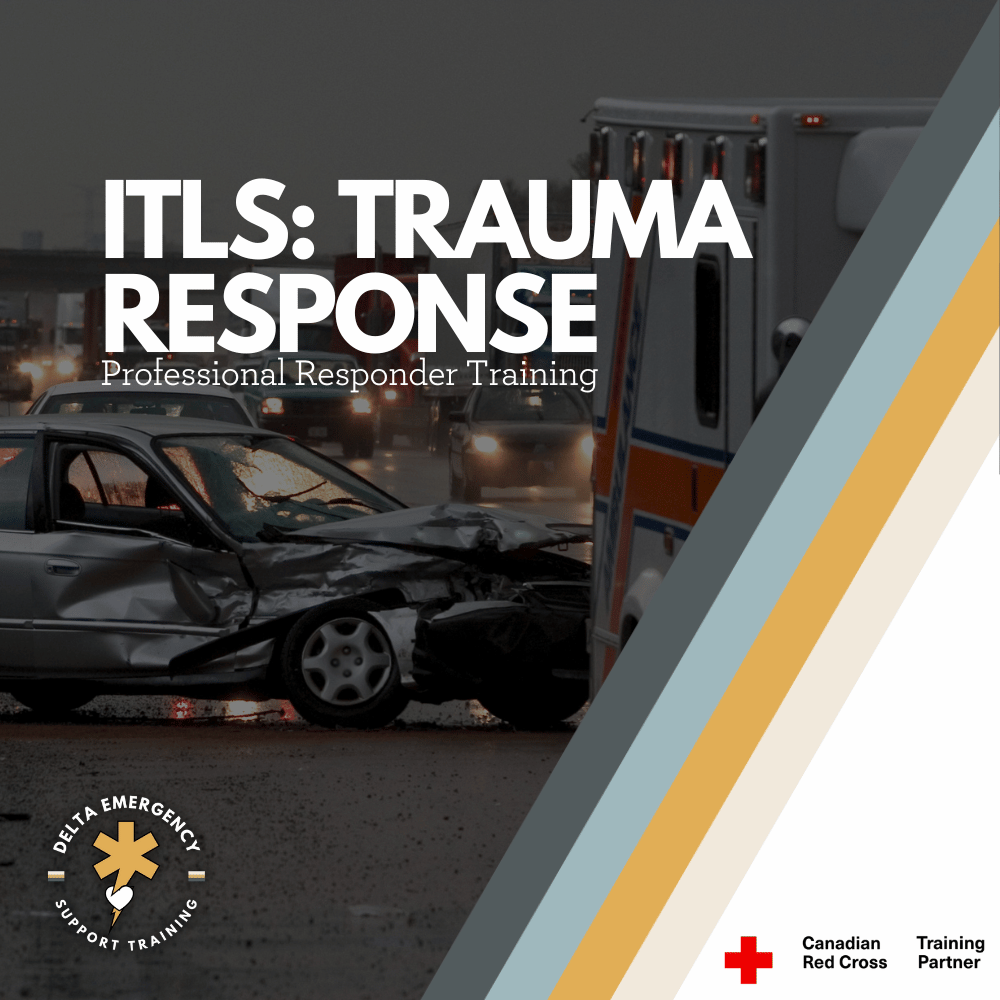
International Trauma Life Support: A Structural Approach to Trauma

Need to take ITLS but not sure what it entails? We’ll break it down for you. International Trauma Life Support (ITLS) gives professional responders the tools to quickly assess, stabilize, and manage trauma patients in high-stress situations. From airway management to bleeding control and spinal care, this course equips you with practical, hands-on skills that could save lives when every second counts.

When emergencies happen, paramedics, firefighters, and advanced first aid responders, having the right tools and knowledge can make the difference between life and death. International Trauma Life Support (ITLS) is a specialized training program that equips professional responders with the skills to rapidly assess, stabilize, and manage trauma patients in high-pressure situations.

If you’re a responder required to take ITLS, here’s what you need to know.

The Purpose of ITLS

ITLS focuses on a structured approach to trauma care. The goal is to give responders a proven framework for:

  • Quickly identifying life-threatening injuries.

  • Performing rapid, effective interventions.

  • Preventing further harm while preparing for transport.

  • Working within professional guidelines and medical protocols.

This training bridges the gap between classroom learning and the unpredictable realities of trauma scenes.

Core Learning in ITLS

Here’s a breakdown of the key skill areas covered in ITLS and what they mean in practice.

1. Patient Assessment & Initial Survey

Responders learn a systematic way to approach every trauma patient:

  • Hazards, Environment, MOI, Patient count and Assistance.

  • Primary survey (C-Spine, LOC, ABCs, RBS,).

  • Identifying life threats in the first few minutes.

Why it matters: A structured assessment ensures nothing is missed under pressure.

2. Airway Management & Adjuncts

Maintaining an open airway is priority number one. ITLS covers:

  • Manual maneuvers (jaw thrust, head-tilt chin-lift).

  • Airway adjuncts (OPA, NPA).

  • Advanced options (supraglottic airways, endotracheal intubation where permitted).

  • Recognizing and managing obstructions.

Why it matters: Without a clear airway, nothing else matters — oxygen can’t reach the body.

3. Breathing & Ventilation Support

Responders are trained to:

  • Recognize respiratory distress and failure.

  • Provide rescue breaths and ventilations with a bag-valve mask.

  • Use oxygen.

  • Identify and treat life-threatening chest injuries (tension pneumothorax, sucking chest wound).

Why it matters: Oxygen delivery is essential to survival, recognizing and treating chest trauma is lifesaving.

4. Circulation & Hemorrhage Control

Stopping major bleeding and supporting circulation includes:

  • Direct pressure and wound packing.

  • Tourniquet application.

  • Recognizing shock and initiating care.

  • Intravenous (IV) or intraosseous (IO) fluid administration (where scope allows).

Why it matters: Uncontrolled bleeding is one of the top causes of preventable trauma deaths.

5. Spinal Motion Restriction (SMR)

Trauma often comes with potential spinal injuries. ITLS emphasizes:

  • Patient assessment for spinal risk.

  • Manual stabilization and cervical collars.

  • Log rolls and backboard use.

  • Updated evidence-based approaches to SMR.

Why it matters: Proper handling prevents permanent paralysis from secondary injury.

6. Head & Chest Trauma Management

Specific scenarios covered include:

  • Managing traumatic brain injuries and increased intracranial pressure.

  • Recognizing chest trauma (flail chest, open chest wounds).

  • Interventions like occlusive dressings, fracture stabilization and deadly bleed control.

Why it matters: These injuries are time-sensitive and require immediate, precise intervention.

7. Protocols & Teamwork

Beyond individual skills, ITLS teaches responders to:

  • Work under medical protocols and established guidelines.

  • Communicate effectively in teams.

  • Prioritize transport decisions (“load and go” vs. “stay and play”).

Why it matters: Trauma care is never solo — effective teamwork saves lives.

Why Professional Responders Need ITLS

For those moving into advanced roles (Advanced First Aid, EMR, PCP, paramedic), ITLS is often a required certification. It ensures all responders use the same language, protocols, and structured assessment, which makes coordination seamless during emergencies.

With ITLS, you’re not just checking a box — you’re developing:

  • Confidence in high-stress scenarios.

  • Competence in trauma management skills.

  • Credibility as a trained professional who can handle life-threatening situations.

Final Takeaway

If you’re required to take ITLS, think of it as more than a course — it’s a survival toolkit for the real world of trauma response. You’ll learn how to rapidly assess patients, manage airways, stop life-threatening bleeds, stabilize spines, and follow trauma protocols effectively.

For professional responders, ITLS is not just training. It’s a commitment to ensuring that when seconds count, you can act decisively, safely, and in line with the best trauma care standards worldwide.

Read More

Scene Assessment for AFA & EMR: A Step-by-Step Guide

Discover how first responders assess scenes in seconds using real tools like HEMPPA, PWCATS, and SCORTS. We teach it in our EMR course — with real-world scenarios to make it stick.

In emergency medical care, scene assessment is your first critical move. It ensures your safety, guides your approach to the patient, and sets the foundation for triage, treatment, and transport. Whether you’re first on the scene or part of a larger response team, a systematic assessment process helps you stay focused and effective.

🚨 Step 1: Scene Overview – HEMPPA

The acronym HEMPPA helps responders quickly assess for overall scene safety and initial priorities:

H – Hazards

  • Look for anything that could harm you, the patient, or bystanders.

  • Examples: traffic, fire, electricity, gas, violence, sharp objects.

  • Why it matters: A dead rescuer helps no one. Always protect yourself first.

E – Environment

  • Assess conditions like weather, lighting, terrain, and location access.

  • Slippery roads, tight spaces, or poor visibility can impact patient care and evacuation.

M – Mechanism of Injury (MOI) / Nature of Illness (NOI)

  • MOI: What caused the injury? (e.g., fall from height, motor vehicle crash)

  • NOI: For medical calls, what’s wrong? (e.g., chest pain, shortness of breath)

P – Patients (Number)

  • Are there multiple patients?

  • Call for more resources if needed (e.g., MVC with multiple occupants).

P – PPE (Personal Protective Equipment)

  • Gloves, eye protection, masks — wear what’s appropriate to protect from bodily fluids or contaminants.

A – Additional Resources

  • Fire, police, ALS units, specialized rescue — ask early.

  • Helps control the scene, provide support, or manage large-scale incidents.

🧠 Step 2: What Else Should You Ask or Observe?

Here are key scene-based questions that build context before you even touch the patient:

  • Position of the patient: Supine, seated, slumped, or ejected from vehicle?

  • Witness reports: What did bystanders or family see? Events leading up to the incident?

  • Chief complaint: What is bothering the patient most right now?

  • Age & gender: Helps form medical differentials quickly.

  • Is the patient tracking/responding? (Eye contact, awareness)

  • Skin colour and condition: Pale, cyanotic, flushed? Cool, clammy, hot? Clues for perfusion.

🔤 PWCATS: For Immediate Patient Impressions

PWCATS helps EMRs gather critical info during the primary assessment.

P – Position

  • Are they standing, lying, slumped, or fetal?

  • Can indicate level of distress or unconsciousness.

W – Witnesses

  • Ask what happened, timing, onset of symptoms, seizure activity, etc.

  • Helps reconstruct events and guide priorities.

C – Chief Complaint

  • What’s their main issue? Chest pain, difficulty breathing, pain scale?

  • Directs your initial focus and treatment.

A – Age

  • Children and elderly present differently.

  • Age affects vital signs, resilience, and urgency of care.

T – Tracking (eye contact/alertness)

  • Are they focused? Following your movements?

  • Quick check of neurologic status or altered mental state.

S – Skin (colour, condition, temperature)

  • Pale, cool, clammy = shock.

  • Flushed, hot = fever or heat stroke.

  • Cyanosis (blue lips/nails) = hypoxia.

⚠️ SCORTS: Scene-Specific Red Flags

Use SCORTS to identify serious, time-sensitive conditions or complicating factors:

S – Stridor / SOB (Shortness of Breath)

  • Upper airway obstruction or respiratory distress?

  • High priority for airway intervention or oxygenation.

C – C-Spine

  • Suspect spinal injury if trauma involved (falls, MVCs).

  • Apply manual stabilization early if indicated.

O – Obvious Bleeding

  • Life-threatening external bleeds need immediate control — pressure, tourniquet, or dressing.

R – Rural / Remote

  • Consider access to definitive care, transport time, or need for air medevac.

  • Distance may increase urgency for certain interventions.

T – Time from ER

  • How far are you from advanced care?

  • May influence treatment decisions, especially in critical cases.

S – Stable / Unstable

  • Is the patient deteriorating? ABCs intact?

  • Helps you determine the transport priority and whether to initiate rapid extrication.

🧭 Scene Assessment in Action: Example Workflow

  1. Pull up, pause, and scan: Use HEMPPA.

  2. Approach safely with PPE on.

  3. Identify number of patients, call for help if needed.

  4. Walk up, observe PWCATS.

  5. Start ABC assessment and initiate any urgent interventions.

  6. Consider SCORTS: look for red flags or complicating factors.

  7. Report findings and updates using structured handover (e.g., ISBAR or CHART).

📋 Summary Cheat Sheet

AcronymStands ForPurpose HEMPPA Hazards, Environment, MOI/NOI, Patients, PPE, Additional ResourcesScene safety & setup PWCATSPosition, Witness, Chief Complaint, Age, Tracking, SkinInitial impression & focused questioning SCORTS Stridor/SOB, C-Spine, Obvious Bleeding, Rural, Time from ER, Stability Red flags & transport factors

🧑‍🏫 Learn Scene Assessment with Us

We teach all of this — and more — in our AFA and EMR classes. You'll learn to apply these acronyms like HEMPPA, PWCATS, and SCORTS in realistic emergency scenarios, helping you build the confidence to assess any scene quickly and safely. Scene assessment is one of the most critical skills a first responder can have, and we make sure you understand why it matters — not just in theory, but in action.

Read More

Pulse Rates and How to Assess Them: Essential Skills for EMR and AFA Students

Understanding how to assess a patient's pulse is a fundamental skill for anyone pursuing EMR or AFA certification. In this blog, we cover everything you need to know about checking pulse rates, including normal ranges for adults, children, and infants, how to locate key pulse points, and how to accurately assess pulse rhythm and quality. Learn about tachycardia, bradycardia, and the significance of a weak or absent pulse, especially in trauma situations.

Understanding Pulse and Heart Rate: Key Concepts for EMRs and AFA Students

In emergency medical response (EMR) or advanced first aid (AFA), one of the essential skills you will learn is how to assess a patient's pulse or heart rate. A person's pulse provides valuable information about their circulatory status, and understanding how to measure it accurately is crucial for diagnosing and managing a variety of medical conditions.

This blog will guide you through the basics of pulse and heart rate, including normal rates for children, adults, and infants. We’ll also dive into how to accurately check a pulse, how to interpret its rhythm and quality, and the significance of an absent or weak pulse, especially in cases of trauma.

What is Pulse?

The pulse is the rhythmic expansion and contraction of an artery as blood is pumped through it by the heart. This beat is felt in various areas of the body known as pulse points. The pulse gives critical information about heart rate, rhythm, and strength.

Normal Pulse Rates by Age

It’s important to know the typical pulse rates for different age groups. The rate can vary based on the individual’s age, activity level, and overall health. Here’s a breakdown of normal resting pulse rates:

  • Infants (0-1 year): 100-160 beats per minute (bpm)

  • Children (1-10 years): 70-120 bpm

  • Adults (11+ years): 60-100 bpm

  • Well-conditioned Athletes: 40-60 bpm (can be normal for some athletes due to a lower resting heart rate)

As you become more familiar with pulse assessment during your EMR or AFA training, you’ll quickly learn how to identify what’s normal for your patient population.

Main Pulse Points and How to Check a Pulse Accurately

As an EMR or AFA student, knowing where to check for a pulse is a fundamental skill. You’ll need to be able to check for a pulse at several key points on the body. Here are the primary pulse points, along with instructions on how to check them accurately:

  1. Radial Pulse (Wrist)

    • Location: On the thumb side of the wrist.

    • How to Check: Place your index and middle fingers on the patient’s wrist, just below the base of the thumb. Apply gentle pressure until you feel the pulse.

    • Position: The patient should be seated or lying down in a relaxed position. It is important not to use your thumb, as it has its own pulse that could interfere with the assessment.

  2. Carotid Pulse (Neck)

    • Location: In the groove of the neck, just to the side of the windpipe.

    • How to Check: Place your index and middle fingers on the side of the patient’s neck, between the trachea and the muscle. Do not press too hard, as it can cause discomfort or interfere with blood flow.

    • Position: The patient can be seated or lying down. If the patient is unresponsive, the carotid pulse is typically the first point to check in an emergency.

  3. Brachial Pulse (Upper Arm)

    • Location: On the inner side of the upper arm, near the elbow.

    • How to Check: Place your index and middle fingers on the inside of the patient’s arm, midway between the shoulder and elbow. Apply gentle pressure to feel the pulse.

    • Position: This pulse is often used to assess infants, as it’s the most reliable pulse point in children under one year old.

  4. Femoral Pulse (Groin)

    • Location: In the crease of the groin, just below the inguinal ligament.

    • How to Check: Place your fingers on the upper thigh, near the groin. Apply firm but gentle pressure to detect the pulse.

    • Position: The patient should be lying down, preferably in a supine position. This pulse is often used in cases of severe trauma or shock.

  5. Posterior Tibial Pulse (Ankle)

    • Location: Behind the medial malleolus (the bony prominence on the inner side of the ankle).

    • How to Check: Place your fingers behind the ankle bone and press gently to feel the pulse.

    • Position: The patient should be lying or sitting with the foot relaxed.

  6. Dorsalis Pedis Pulse (Foot)

    • Location: On the top of the foot, between the first and second metatarsals.

    • How to Check: Place your fingers on the top of the foot, slightly to the medial side, and gently press to feel the pulse.

    • Position: The patient should be in a seated or supine position, with the foot relaxed.

How to Assess Pulse Rhythm and Quality

When assessing a pulse, it’s essential to evaluate not only the rate but also the rhythm and quality:

  • Rhythm: The rhythm of the pulse refers to the regularity of the heartbeat. It can be classified as:

    • Regular: The pulse follows a consistent, steady pattern.

    • Irregular: The pulse has an erratic or uneven rhythm, which could indicate an arrhythmia or other heart conditions.

  • Quality: The quality of the pulse refers to its strength. A pulse can be:

    • Strong and Bounding: A pulse that is easy to feel and forceful, which could indicate conditions such as fever, anxiety, or hyperthyroidism.

    • Weak or Thready: A pulse that is faint and difficult to feel, often indicating shock, blood loss, or dehydration.

    • Absent: If no pulse is felt at a pulse point, it may suggest severe trauma, a blocked artery, or cardiac arrest.

Tachycardia and Bradycardia

When evaluating pulse, you may encounter conditions such as tachycardia (a rapid heart rate) or bradycardia (a slow heart rate):

  • Tachycardia: A heart rate that exceeds 100 bpm in adults. Tachycardia can be caused by fever, stress, anemia, or cardiovascular problems.

  • Bradycardia: A heart rate below 60 bpm in adults. Bradycardia can indicate heart conduction issues, but it is also common in well-conditioned athletes.

Weak or Absent Pulse in Trauma

In trauma cases, an absent or weak pulse can be a critical sign. Trauma, especially internal bleeding or blocked arteries, can lead to a diminished pulse. If there is significant internal hemorrhaging, the pulse may become weaker in the lower extremities or other areas far from the heart. In cases of severe blood loss, the body shunts blood away from the extremities to maintain circulation to vital organs, resulting in weak or absent pulses in the arms or legs.

A blocked artery due to a clot, injury, or constriction can also result in an absent pulse distal to the blockage. Immediate intervention is needed in these cases to restore blood flow and stabilize the patient.

Conclusion

As an EMR or AFA student, learning to assess a patient’s pulse accurately is essential to providing effective care in emergency situations. Understanding the normal pulse rates, knowing how to check a pulse at key locations, and being able to interpret pulse rhythm and quality will greatly improve your ability to assess a patient’s condition. Additionally, recognizing the significance of weak, absent, or irregular pulses can help you identify life-threatening conditions like trauma, internal bleeding, or blocked arteries.

At Delta Emergency, we teach our students how to properly assess and respond to real-life emergencies. With expert instructors who are experienced paramedics, our courses ensure that you're prepared to make quick and informed decisions during critical situations.

Read More