The Delta Dispatch
Understanding Basic, Intermediate, and Advanced Airway Adjuncts in Prehospital Care
Airway management is the cornerstone of emergency care. From simple oropharyngeal and nasopharyngeal airways to supraglottic devices and endotracheal intubation, prehospital providers need to know when—and how—to use each tool. This guide explains the essential skills and decision-making strategies for basic, intermediate, and advanced airway adjuncts to help responders keep patients breathing and safe.
Securing and maintaining a patient’s airway is the cornerstone of prehospital medicine. Whether you are an Emergency Medical Responder (EMR), Primary Care Paramedic (PCP), or Advanced Care Paramedic (ACP), understanding the range of airway adjuncts—and when to use them—is critical for optimal patient outcomes. Airway adjuncts are commonly grouped into three categories: basic, intermediate, and advanced.
1. Basic Airway Adjuncts
These devices are non-invasive and require minimal training, yet they can be lifesaving.
Oropharyngeal Airway (OPA)
Use: Unconscious patients without a gag reflex to keep the tongue from obstructing the airway.
Key Points: Measure from the corner of the mouth to the angle of the jaw; insert upside down then rotate 180° (or sideways with a tongue depressor in children).Nasopharyngeal Airway (NPA)
Use: Patients with an intact gag reflex or trismus (jaw clenching).
Key Points: Lubricate well; size from nostril to earlobe. Contraindicated in suspected basilar skull fractures.Suction Equipment
Use: Clears secretions, blood, or vomit to maintain a patent airway.
Clinical Pearl: Basic adjuncts buy time. Always reassess and be ready to escalate if ventilation or oxygenation remains inadequate.
2. Intermediate Airway Adjuncts
These devices provide more definitive control without requiring endotracheal intubation skills.
Supraglottic Airway Devices (SADs) such as:
Laryngeal Mask Airway (LMA)
King LT or i-gel
Use: For unconscious patients when bag-valve-mask (BVM) ventilation is inadequate or prolonged transport is expected.
Key Points: Rapid insertion, minimal training compared to intubation, and useful when laryngoscopy is not feasible.
3. Advanced Airway Adjuncts
These procedures require specialized training and are often reserved for paramedics with advanced certification or physicians.
Endotracheal Intubation (ETI)
Use: To provide definitive airway protection and control ventilation.
Key Points: Requires laryngoscopy, confirmation of tube placement (capnography is gold standard), and ongoing monitoring for dislodgement.Surgical Airway (Cricothyrotomy or Needle Cricothyrotomy)
Use: “Cannot ventilate, cannot intubate” scenarios such as severe facial trauma or airway obstruction.
Key Points: High-stakes, last-resort procedure with strict indications.
Airway Decision-Making in the Field
When deciding which airway adjunct to use, consider:
Level of provider certification and local protocols.
Patient condition: Consciousness, gag reflex, trauma, and potential for rapid deterioration.
Environment: Limited space, lighting, or access may guide your choice.
Tip: Airway management is dynamic. Start with the least invasive method and escalate as needed while continually reassessing breathing and oxygenation.
Training and Maintenance
Regular Practice: Skills such as BVM ventilation and intubation degrade quickly without use.
Equipment Checks: Verify availability, integrity, and proper sizes of adjuncts at the start of every shift.
Bottom Line
From OPAs and NPAs to supraglottic devices and endotracheal tubes, airway adjuncts form a spectrum of tools for professional responders. Mastery of their indications, insertion techniques, and limitations ensures that you can match the right device to the right patient, improving survival and reducing complications in the prehospital setting.
Hypoxia 101: Symptoms, Causes, and First Aid Tips
Hypoxia can lead to irreversible brain damage within minutes. Rapid recognition, airway management, aggressive oxygenation, and swift treatment of the underlying cause—especially opioid-related respiratory depression—are essential to preserving neurological function and improving outcomes.
Oxygen is the brain’s primary fuel, and even a short interruption can trigger irreversible injury. Understanding how hypoxia develops—and staying current with evolving science—is critical for anyone providing emergency medical care.
Current Causes of Hypoxia
Hypoxia can result from a wide range of emergencies, with some trends growing more prominent:
Opioid Overdose: Canadian surveillance data through 2024 show that potent synthetic opioids (including illicit fentanyl analogues) are driving an increase in severe respiratory depression and sudden apnea.
Respiratory Compromise: Severe asthma, COPD exacerbations, airway obstruction, and anaphylaxis remain frequent triggers. Climate-related wildfire smoke has also increased smoke-inhalation hypoxia incidents.
Circulatory Emergencies: Cardiac arrest, massive hemorrhage, and shock reduce perfusion and oxygen delivery to the brain.
Brain Pathophysiology: What’s New
Recent neuroimaging research highlights how quickly hypoxia damages neurons:
First 1–2 Minutes: Early synaptic failure leads to measurable cognitive impairment even before loss of consciousness.
4–6 Minutes: MRI studies confirm the hippocampus—essential for memory—is especially vulnerable, making early intervention vital for long-term cognitive outcomes.
Beyond 6 Minutes: Widespread cortical cell death often leads to permanent neurologic deficits.
Key Clinical Indicators
Altered mental status: confusion, agitation, sudden lethargy
Central cyanosis of lips or tongue
Hypoventilation or apnea, particularly in opioid overdose
SpO₂ decline despite supplemental oxygen, suggesting perfusion or carbon monoxide issues
Seizure activity or abnormal pupillary responses
Field Management Priorities
Airway & Ventilation
Rapid airway assessment and maintenance with OPA/NPA or supraglottic airway as indicated.
Bag-valve-mask ventilation if hypoventilation or apnea is present.
Oxygenation
Deliver high-concentration oxygen via the appropriate device.
Monitor with pulse oximetry and end-tidal CO₂ for early detection of ventilation problems—portable capnography is now standard in many Canadian ambulances.
Cause-Specific Interventions
Opioid Overdose: Administer naloxone per protocol while providing ventilatory support. Early naloxone use significantly reduces hypoxic brain injury.
Asthma/COPD: Supportive bronchodilator therapy if within scope.
Shock or Hemorrhage: Rapid bleeding control and perfusion support.
Neurologic Monitoring
Frequent Glasgow Coma Scale checks, pupillary assessments, and continuous reassessment during transport or care.
New Guidance Highlights
Avoid Hyperoxia: Updated resuscitation guidelines emphasize maintaining SpO₂ between 94–98% to prevent oxygen toxicity.
Delayed Effects: Evidence shows even brief hypoxia can lead to delayed neurocognitive changes, underscoring the importance of early recognition and thorough documentation for hospital teams.
Simulation Training: Advanced simulation with real-time capnography is increasingly used in training programs to prepare responders to identify subtle respiratory decline.
Key Take away Hypoxia can lead to irreversible brain damage within minutes. Rapid recognition, airway management, aggressive oxygenation, and swift treatment of the underlying cause—especially opioid-related respiratory depression—are essential to preserving neurological function and improving outcomes.
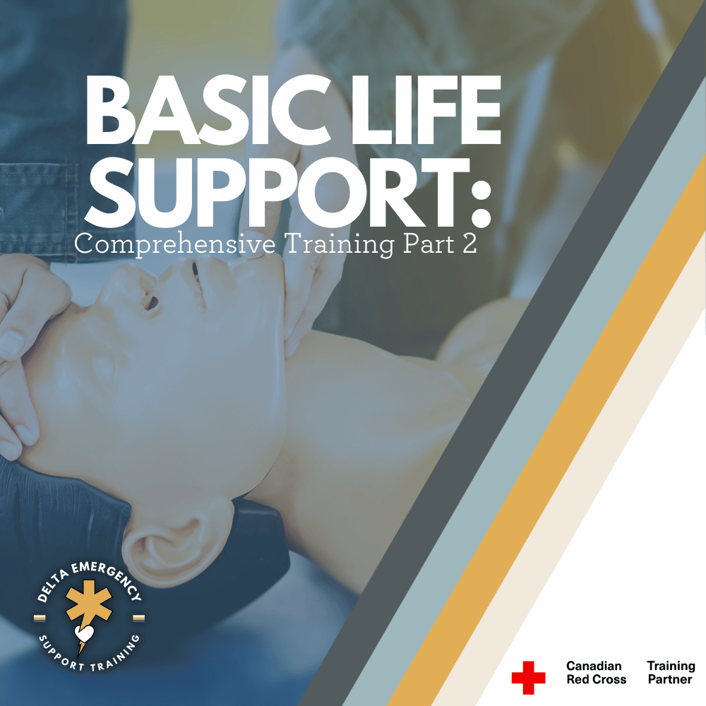
Basic Life Support Training Series: Part 2
Are you prepared to save lives during emergencies? Mastering Basic Life Support (BLS) is crucial, and understanding the key steps for assessing airway, breathing, and circulation is essential. Discover effective techniques like the head tilt-chin lift and jaw thrust for opening the airway, and learn about devices such as oropharyngeal airways (OPA) and nasal cannulas for maintaining clear breathing passages. Find out how to assess circulation through pulse checks, capillary refill, and extremity warmth. Explore these vital BLS skills and be ready to make a difference when it matters most. Boost your life-saving capabilities now!
Welcome back to the second part of our blog series on Basic Life Support (BLS). In the first blog, we discussed scene assessment, the ABC check (Airway, Breathing, and Circulation), and the proper use of CPR and AED. Now, in part two, we will delve into life-saving interventions and devices used within each step of the ABC check. Let's explore the crucial steps that can make a significant difference in saving lives.
Head tilt chin lift in basic life support training class
Life Saving Interventions: Airway
Airway management is a critical component of Basic Life Support (BLS) and plays a crucial role in ensuring the patient's ability to breathe effectively. In addition to the manual maneuvers we discussed earlier, such as the head tilt-chin lift and jaw thrust, there are other devices that can be used to secure and protect the airway. Let's explore two commonly used devices: the Oropharyngeal Airway (OPA) and the Nasopharyngeal Airway (NPA).
Oropharyngeal Airway (OPA):
The oropharyngeal airway, is a curved plastic tube that is inserted into the patient's mouth to maintain the patency of the airway. It serves two main purposes:
a) Clearing Obstructions: The OPA helps to displace the tongue from the back of the throat, preventing it from obstructing the airway and impeding the passage of air. This is used for unconscious patients or those with decreased consciousness.
b) Facilitating Manual Stabilization: By providing a firm structure, the OPA allows rescuers to perform manual stabilization of the patient's head and neck, as it helps maintain the alignment of the airway.
When using an OPA, it is important to select the correct size for the patient to ensure proper placement and prevent complications. Insert the device upside down, rotating it 180 degrees as it reaches the back of the throat. Proper placement should allow the device to sit between the teeth or lips and the base of the tongue.
Nasopharyngeal Airway (NPA):
The nasopharyngeal airway, or nasal airway, is a flexible tube inserted through the nostril into the back of the throat. It is particularly useful in patients who have an intact gag reflex or clenched teeth, making oral insertion difficult or risky. The NPA has several advantages:
1) Improved Comfort: The NPA is generally better tolerated by conscious or semi-conscious patients as it avoids stimulation of the gag reflex.
2) Unobstructed Oral Access: By securing the airway through the nasal passage, the mouth remains free and accessible for other procedures, such as suctioning or oral medication administration.
To insert an NPA, select the appropriate size by measuring from the tip of the nostril to the earlobe or the angle of the jaw. Lubricate the device before gently inserting it along the floor of the nasal passage, aiming towards the back of the throat. Monitor for signs of discomfort or obstruction during insertion and adjust if necessary.
It is important to note that the use of OPAs and NPAs requires proper training and an understanding of the patient's anatomy and potential contraindications. Rescuers should be cautious and attentive to the patient's response while using these devices.
By employing manual maneuvers like the head tilt-chin lift and jaw thrust, along with the use of OPAs and NPAs, rescuers can effectively secure and maintain an open airway for patients in need of Basic Life Support. Remember, accurate assessment and regular practice are essential to ensure proper airway management in emergency situations.
Life Saving Interventions: Breathing
Various devices can be used to provide oxygenation and ventilation support. Let's explore the different devices commonly used in BLS:
Nasal Cannula:
The nasal cannula is a lightweight device that delivers supplemental oxygen through two small prongs inserted into the patient's nostrils. It provides a low to moderate flow rate of 1-4 liters per minute (LPM) with an oxygen concentration ranging from 25% to 45%. The nasal cannula is comfortable and allows patients to speak, and move freely while receiving oxygen.
Simple Face Mask:
The simple face mask is a mask that covers the patient's nose and mouth, delivering oxygen at a flow rate of 6-10 LPM. It provides a higher oxygen concentration compared to the nasal cannula, ranging from 40% to 60%. The simple face mask is commonly used in patients who require higher oxygen levels or when a more precise oxygen concentration is not required. However, it may impede communication and patient comfort.
Non-Rebreather:
The non-rebreather is a mask with a reservoir bag attached, ensuring the delivery of high concentrations of oxygen. It has one-way valves that prevent the patient from inhaling exhaled air and allow for a flow rate of 10-15 LPM. The non-rebreather mask can deliver an oxygen concentration of 90% or higher. It is particularly useful in patients with severe hypoxia or in cases where precise oxygen titration is necessary.
Bag-Valve-Mask (BVM):
The bag-valve-mask (BVM) device, is a manually operated resuscitator used to provide positive pressure ventilation. It consists of a self-inflating bag, a one-way valve, and a mask. The BVM is connected to an oxygen tank with a flow rate of 15 LPM, providing an oxygen concentration close to 100%. The BVM is often used in cases of respiratory distress or respiratory arrest, when the patient is not breathing adequately or requires assisted ventilation.
During assisted ventilation with a BVM, 1/3 of the bag should be compressed once every 5-6 seconds to achieve a target ventilation rate of 10-12 breaths per minute.
Newer BVM’s include a safety valve that makes a noise if you are compressing the bag too hard or fast, allowing for user friendly use.
As you provide assisted ventilation using a BVM, pay close attention to the patient's response. Look for chest rise and fall with each breath delivered. The presence of chest rise indicates that the ventilation is effectively reaching the patient's lungs.
Additionally, observe the patient's overall color. Effective ventilation should help improve the patient's skin color, with a return to a more normal or healthier appearance. If the patient's color improves, it is a positive sign that oxygenation and ventilation are being adequately supported.
It is important to note that when administering oxygen, the flow rate should be carefully adjusted based on the patient's condition and oxygen saturation levels. Oxygen therapy should be monitored closely.
Circulation assessment
Circulation assessment is a crucial step in Basic Life Support (BLS) to determine the adequacy of blood flow. While CPR remains the life-saving intervention when a pulse is not detected, let's explore other methods to assess circulation when a pulse is present.
Capillary Refill: To assess capillary refill, gently press down on the patient's nail bed or the tip of their finger. Upon releasing the pressure, observe how quickly the color returns to the area. A normal capillary refill time is around 2 seconds. If color returns within this timeframe, it indicates good capillary refill and suggests adequate peripheral circulation.
Extremity Warmth: Feeling the temperature of the patient's extremities, such as their hands and feet, can provide valuable information about circulation. Warm extremities indicate good blood flow, while cold extremities may suggest impaired circulation.
It's important to note that these assessments are complementary to the presence of a pulse. If there are concerns about circulation or any signs of poor perfusion, prompt medical attention should be sought.
Circulatory Obstruction: Major blood loss from an injury can obstruct circulation and pose a life-threatening situation. In such cases, immediate action is crucial. If the hemorrhage is external, apply direct pressure to the bleeding site using PPE always. Maintaining pressure helps control the bleeding and promotes clot formation, thereby restoring circulation. If available, consider the use of a tourniquet for severe hemorrhage that cannot be controlled by direct pressure alone.
Remember, it is important to prioritize personal safety and seek professional medical help as soon as possible in cases of significant bleeding or other circulatory emergencies. Timely intervention can make a significant difference in patient outcomes.
Training
Mastering Basic Life Support (BLS) is crucial for healthcare professionals, including nurses, doctors, dentists, firefighters, and other first responders. Understanding the key steps for assessing and applying critical interventions for airway, breathing, and circulation is essential to deliver timely and effective care during emergencies. At Delta, we recognize the importance of BLS training, as it is often required to be completed yearly for professionals in various fields. We are dedicated to providing frequent BLS courses to equip individuals with the life-saving skills they need. Enroll in our BLS courses today and stay prepared to make a difference when it matters most.
Firefighter Training: OPA's and NPA's for Airway Management
Nasopharyngeal airways (NPA) and oropharyngeal airways (OPA) are commonly used in healthcare settings to maintain a patient's airway and assist with breathing.When used correctly, NPAs can help clients who may be experiencing respiratory distress during certain treatments. However, it's important to have the necessary knowledge and protocols in place to safely insert and monitor the device. At Delta Emergency Support Training, we can help you learn how to safely use NPAs in spa settings and beyond. Our training sessions include a range of courses, including Standard First Aid (SFA), Advanced First Aid (AFA), and Emergency Medical Responder (EMR), and we offer in-person, hybrid, and online options to suit different needs. Contact us at info@deltaemergency.com to learn more about our training sessions and how we can help you develop the skills and knowledge you need to respond to emergency situations effectively.
As a firefighter, you need to be prepared to respond to emergency situations, including those involving airway management for an unconscious patients airway. Two commonly used tools for airway management are oropharyngeal airways (OPA's) and nasopharyngeal airways (NPA's). Today we'll discuss what these devices are, why they are used, and how to properly insert them as a first responder.
Four colour coded Oropharyngeal airways lay across a medical sheet.
What are OPA's and NPA's?
Oropharyngeal airways (OPA's) and nasopharyngeal airways (NPA's) are medical devices used to keep an airway open in patients who can’t secure it themselves. They are used when the patient is unable to maintain their own airway due to injury, illness, or drug overdose and become unconscious. OPA's and NPA's are both designed to be inserted into the patient's airway to help keep it open.
An OPA is a curved plastic device that is inserted into the patient's mouth and extends into the pharynx. It helps to prevent the tongue from obstructing the airway and allows air to pass freely into the lungs. OPA's are available in different sizes to accommodate patients of different ages and sizes.
An NPA is a flexible, hollow tube that is inserted into the patient's nostril and extends into the nasopharynx. It helps to keep the airway open and can be used in patients who are unable to tolerate an OPA due to gag reflex or other reasons.
Why do we use OPA's and NPA's?
The primary reason for using OPA's and NPA's is to maintain a patient's airway and ensure that they are able to breathe. When a patient's airway is obstructed, it can quickly lead to hypoxia, which is a dangerous condition where the body's tissues do not receive enough oxygen. OPA's and NPA's are used to prevent this from happening by keeping the airway open and allowing oxygen to flow freely into the lungs.
Testing for LOC (Level Of Consciousness)
Before inserting an OPA, it is important to assess the patient's level of consciousness. The AVPU scale is a simple and quick method for assessing a patient's level of consciousness based on their response to stimuli. The scale has four categories:
A: Alert
V: Verbal response
P: Pain response
U: Unresponsive
To use the AVPU scale, a healthcare provider would first try to get the patient's attention by speaking to them in a normal tone of voice. If the patient responds appropriately, they would be classified as "A" for alert. If the patient responds to verbal stimuli but is not fully alert, they would be classified as "V" for verbal response. If the patient only responds to painful/physical stimuli, such as a trap squeeze or nail bed pressure, they would be classified as "P" for pain/physical response. If the patient does not respond to any stimuli, they would be classified as "U" for unresponsive.
If a patient is assessed as being unresponsive or only responding to painful stimuli, an OPA may need to be inserted to maintain their airway. However, if the patient is alert or responding to verbal stimuli, an OPA may not be necessary.
How to insert OPA's and NPA's as a first responder
Proper insertion of OPA's and NPA's is crucial to their effectiveness and patient safety. Here are some general steps for inserting OPA's and NPA's:
Inserting an OPA:
Choose the appropriate size OPA for the patient based on their age and size. Measure from the corner of the mouth to the angle of the jaw.
Position the patient's head in a neutral position.
Open the patient's mouth using the cross finger technique.
Insert the OPA into the patient's mouth, with the curved end facing the roof of the mouth.
Gently rotate the device 180 degrees as you insert it, until it is in a anatomical position.
Inserting an NPA:
Choose the appropriate size NPA for the patient based on their age and size by measuring the tip of the nose to the corner of the jaw.
Lubricate the NPA with a water-soluble lubricant.
Position the patient's head in a neutral position.
Insert the NPA into the patient's nostril starting with the right nostril. Bevelled edge facing the septum.
Gently advance the device until it reaches the back of the nasopharynx
At Delta Emergency Support Training, we understand the importance of being prepared for medical emergencies. We offer a range of training sessions to help individuals and classes develop the skills and knowledge they need to respond to emergency situations. Our courses include Standard First Aid (SFA), Advanced First Aid (AFA), and Emergency Medical Responder (EMR), and we offer in-person, hybrid, and online options to suit different needs.
If you have any questions about our training sessions or medical emergencies in general, please don't hesitate to reach out to us. You can contact us at info@deltaemergency.com and we'll be happy to assist you.
What is an EMR?
The first step for an EMR is to assess the situation for hazards that can harm themselves, their partners, and their patients, and determine the nature of the emergency. They need to determine the level of medical attention needed and make quick decisions…
Behind the Scenes of an Emergency Medical Responder: Saving Lives with Speed and Skill
An Emergency Medical Responder (EMR) is a healthcare professional who is trained to provide immediate medical care in emergency situations. EMRs are often the first healthcare providers to arrive on the scene of an emergency, and they work closely with other healthcare professionals to ensure that patients receive the appropriate care and treatment. EMRs are trained to provide basic life support measures, including administering CPR, controlling bleeding, treating shock, managing spinal injuries, and stabilizing fractures. They are also trained to manage patients with a variety of medical emergencies, including cardiac arrest, respiratory distress, and trauma. Here are some of the typical job functions of an EMR:
1. Assess the situation:
The first step for an EMR is to assess the situation for hazards that can harm themselves, their partners, and their patients, and determine the nature of the emergency. They need to determine the level of medical attention needed and make quick decisions based on their assessment.
2. Stabilize Patients:
EMRs are trained to stabilize patients by maintaining their airways and providing necessary interventions such as assisted ventilations, medical oxygen, life-threatening bleeding, and medication.
2. Administer First Aid:
EMRs are trained to provide basic-intermediate-advanced first aid such as CPR, wound management, and immobilization of injured patients.
4. Communicate with the medical team:
EMRs communicate vital information to the medical team, such as the patient's vital signs, symptoms, and any other relevant medical history.
5. Provide Emotional Support:
EMRs provide emotional support to patients and their families during times of crisis. They should be compassionate and empathetic, helping to ease the stress and anxiety that often come with medical emergencies.
6. Document the incident:
EMRs must document the incident, including vital signs, patient history, and any medical interventions administered.
7. Transport Patients:
EMRs are responsible for transporting patients to medical facilities safely. Overall, the role of an EMR is crucial in emergency medical situations, and they must be well-trained, quick-thinking, and compassionate individuals. EMRs work in a variety of settings, including ambulance services, fire departments, and hospitals. They play a critical role in the healthcare system by providing immediate medical care to patients in emergency situations, and their actions can often make the difference between life and death.
The road to EMR certification can be confusing. The EMR program is different in provinces like BC, Alberta, Saskatchewan, and Manitoba. The Canadian Red Cross EMR, AFA, or FR curriculum is identical across Canada but provincial standards and must be taught in respective provinces. At Delta Emergency, we understand what certifications you need for your future career, how and where to obtain them, and what type of work will be available to you when you’re fully certified. Reach out with questions!
BVM
Maintain an Airway, use HTCL or MJT, use Suction, insert an OPA or NPA, begin Ventilations and apply Oxygen. BLS, FR, AFA, and EMR
This is a basic and generalized overview of Emergency Medical Responders across North America. When in doubt, follow your local protocols! Contact us if you have any specific questions about the EMR scope of practice and career path in Alberta. We are industry experts and have been working full time in EMS for years.
At Delta Emergency Support Training we are happy to answer any questions you may have about becoming an EMR. We provide training sessions for individuals or classes. We have options for in person, hybrid or online classes for Standard First Aid (SFA) Advanced First Aid (AFA) and Emergency Medical Responder (EMR) courses. For all inquiries please email info@deltaemergency.com

