The Delta Dispatch

SAGER: Traction Splinting A Midline Femur Fracture

At Delta Emergency Support Training, we provide in-depth advanced first aid classes that cover a variety of topics, including the proper application of a Sager splint. Our classes are designed for medical professionals and advanced first aiders who want to expand their knowledge and skills. In this guide, we'll cover how to effectively use a Sager splint to immobilize femoral shaft fractures and other leg injuries.

What is a Sager Splint?

A Sager splint, also known as a traction splint, is a medical device used to immobilize and align a fractured or injured leg. It is designed to reduce pain and swelling by applying traction to the leg, pulling the bone fragments back into alignment. The splint consists of two main components: the splint itself, which is a long, narrow rod that extends from the foot to the groin, and the traction mechanism, which is attached to the foot end of the splint.

An advanced first aider applies a Sager splint to a person's leg.

An advanced first aider applies a Sager splint to a patient with a broken femur. The patient is lying on their back and the first aider is wearing white gloves and kneeling beside them.

The splint is typically made of aluminum or carbon fiber and is lightweight and easy to transport. It is designed to be adjustable to fit a variety of leg sizes and can be quickly and easily applied in emergency situations. The traction mechanism is usually a ratcheting system that allows the medical professional to control the amount of traction being applied to the leg. This mechanism can be adjusted to achieve the desired amount of traction and can be released easily if necessary.

The amount of pressure applied is weighed in pound with a rule of thumb being 10% of the patients body weight for a maximum of 15 pounds per femur fracture or 30 pounds for a bilateral femoral fracture.

Sager splints are used for femoral shaft fractures, which are fractures that occur in the middle of the thigh bone. These types of fractures can be serious and require immediate attention. Sager splints are effective at immobilizing the affected leg, reducing pain and swelling, and realigning the bone fragments.

What causes a femur fracture?

Femur fractures can occur due to a variety of mechanisms of injury (MOI), with high-energy trauma and direct impact being the most common causes.

High-energy trauma, which involves a significant amount of force, can be caused by car accidents, falls from a height, sports injuries, and other incidents that involve significant force. The femur can break due to the impact of the force or due to bending or twisting of the bone.

Direct impact to the thigh bone, such as a blow from a heavy object or a fall onto a hard surface, is another common MOI that can cause a femur fracture. This can occur in industrial accidents or during contact sports. In both cases, the force applied to the femur is greater than the bone's ability to absorb it, causing the bone to break.

It's important to note that the severity of the femur fracture can vary depending on the MOI and the angle of impact.

How serious is a broken femur?

A femur break, particularly a bilateral femur fracture, can be a serious and potentially limb-threatening injury. The femur is the longest and strongest bone in the human body and is responsible for supporting the weight of the body and facilitating movement. When the femur is fractured, it can cause significant pain, swelling, and deformity of the affected leg.

One of the most significant dangers of a femur break is the potential for the femoral artery to be compromised by a bone fragment. The femoral artery is a large blood vessel that runs through the thigh and supplies blood to the lower leg and foot. If a bone fragment from a femur break punctures the femoral artery, it can lead to internal bleeding, which can be life-threatening if not treated promptly.

In addition to the danger of internal bleeding, a femur break can also result in nerve damage, muscle damage, and compartment syndrome. Compartment syndrome occurs when pressure within a muscle compartment builds up, which can cause damage to the nerves, blood vessels, and muscles in the affected area. If left untreated, compartment syndrome can lead to permanent damage or loss of function in the affected limb.

Indications and contraindications

Indications:

  • Femoral shaft fractures:

    Sager splints are commonly used to immobilize and align femoral shaft fractures, which are fractures that occur in the middle of the thigh bone. Bi-lateral or single femur fractures.

Contraindications:

  • Open fractures:

    A compound or open fracture is a type of bone fracture where the broken bone pierces through the skin.

    In a compound fracture, the exposed bone can be seen or felt through the wound in the skin, and there may be visible bleeding, swelling, and/or deformity at the site of the injury. If the skin over the fracture is broken or torn, the Sager splint should not be applied as it can cause further damage or infection.

  • Injuries on the effected leg:

    If the patient has any other associated injuries, such as ankle or fib tib fractures, a Sager splint may be contraindicated. Additionally, if the patient has a knee injury, such as a dislocated knee, a Sager splint may not be appropriate as it can exacerbate the injury. It's important to carefully evaluate the patient with a thorough rapid body survey to rule out injuries that may effect application.

  • Life threatening injuries:

    If a patient has a life-threatening injury, the priority is to rapidly transport the patient to a medical facility for appropriate treatment. In such cases, immobilization with a Sager splint may not be the best course of action, as it can take time to properly apply the splint, and rapid transport may be necessary to save the patient's life.

    It's important to remember that in cases of multiple injuries or life-threatening injuries, the priority is to save the patient's life, life over limb. While a femur fracture can be a serious and potentially limb-threatening injury, other injuries may take precedence over the fracture.

Proper application

Assuming that the patient has been assessed and a Sager splint is deemed appropriate, the medical professional or advanced first aider will follow the steps outlined in the previous response to properly apply the splint. Proper application of a Sager splint requires medical training and should only be done by trained professionals. Here are the general steps involved in applying a Sager splint:

  1. Assessment of the patient's injury:

    Before applying a Sager splint, the medical professional should assess the patient's injury to determine if it's the right tool for the job. A physician should evaluate indications and contraindications.

  2. Patient in a supine position:

    Once it's been determined that a Sager splint is necessary, the patient should be placed in a supine position.

  3. Remove clothing from effected area:

    Any clothing that obstructs the affected leg should be trauma stripped to ensure proper placement of the Sager as well as to check for compound fractures.

  4. Placement of the Sager:

    The Sager splint should be placed on the affected leg, positioning the ankle and foot in the appropriate spot. The splint should be placed with the traction component of the device facing the foot.

  5. Securing the splint to the leg:

    The splint should be secured to the leg with straps or bandages, ensuring that it is snug but not too tight. This will help to keep the splint in place and provide the necessary support.

  6. Application of traction:

    The traction component of the Sager splint should be applied, pulling the leg gently and steadily until the desired amount of traction is achieved. This will help to reduce pain and swelling and realign the bone fragments.

  7. Pulse, sensation, and movement check:

    The medical professional should check the patient's pedal pulse, sensation, and movement in the affected leg to ensure that the splint is not causing further damage or obstruction.

    Training

    Sager splints are orthopedic devices that are commonly used to immobilize femoral shaft fractures. They can help to stabilize the fracture and prevent further damage to the leg or other parts of the body. However, the proper application of a Sager splint requires specialized training and should only be done by trained medical professionals or advanced first aiders.

    At Delta Emergency Support Training, we provide in-depth advanced first aid classes that cover a variety of topics, including the proper application of a Sager splint.

Read More

Treating Chest Pain: Aspirin and Nitroglycerin

Aspirin and nitroglycerin are two medications that are commonly used by first responders to treat chest pain or suspected heart attack. Aspirin is a platelet inhibitor that helps to prevent blood clots from forming and reduces the risk of further damage to the heart. Nitroglycerin is a vasodilator that helps to increase blood flow to the heart by dilating blood vessels, which reduces the workload on the heart muscle.

While these medications can be effective in treating chest pain or suspected heart attack, it's important for first responders to be familiar with their uses, risks, and benefits, and to follow established protocols and guidelines when administering them. Aspirin and nitroglycerin can have potential side effects and contraindications, so it's important to use them only as directed and under the guidance of a healthcare professional.

To ensure the safety and well-being of their patients, first responders should also educate patients about the benefits and risks of aspirin and nitroglycerin, and the importance of seeking medical follow-up after receiving these medications. By working closely with healthcare professionals and following established protocols and guidelines, first responders can provide effective care and help ensure the safety and well-being of their patients.

When it comes to treating chest pain or suspected heart attack, aspirin and nitroglycerin are two medications that first responders may use to help their patients depending on scope of practice. But what are these medications, how do they work, and what do first responders need to know about their use?

man sitting down and clutching his chest in pain. He appears to be experiencing a heart attack. Calgary Fire Fighters on the way.

Aspirin:

What is Aspirin, and How Does it Work?

Aspirin, also known as acetylsalicylic acid (ASA), is a medication that's commonly used to treat pain, fever, and inflammation. Aspirin works by inhibiting the activity of platelets, which are small blood cells that play a key role in blood clotting. When a blood vessel is damaged, platelets are activated and begin to stick to the damaged area to form a plug, which helps to stop bleeding. However, when a blood clot forms inside an artery supplying the heart, it can block blood flow and cause a heart attack.

As a platelet inhibitor, aspirin helps to prevent blood clots from forming and reduces the risk of further damage to the heart.

How is Aspirin Used in Emergency Situations?

Aspirin is an important medication for first responders to use in emergency situations, particularly for patients experiencing chest pain or suspected heart attack also know as a myocardial infarction. To speed up its onset of action, chewing aspirin is often recommended. The recommended dose of aspirin for patients with suspected heart attack is typically between 160-325 mg.

Contraindications:

There are certain situations where aspirin use may not be appropriate or may need to be used with caution.

Some common contraindications for aspirin use include:

  • Allergy or hypersensitivity to aspirin or other non-steroidal anti-inflammatory drugs (NSAIDs)

  • Active bleeding or bleeding disorders

  • Suspected stroke

  • ASA exacerbated asthma

  • Children with viral infection

    Nitroglycerin:

    What is Nitroglycerin, and How Does it Work?

    Nitroglycerin is a medication used to treat chest pain or angina, which is often a symptom of heart disease. Nitroglycerin is a vasodilator, which means that it relaxes and widens blood vessels throughout the body, including the coronary arteries that supply blood to the heart muscle.

    By dilating the blood vessels, nitroglycerin helps to increase blood flow to the heart, which reduces the workload on the heart muscle and can relieve chest pain or angina. Nitroglycerin works by releasing nitric oxide, which is a gas that relaxes the smooth muscle cells in the walls of blood vessels.

    In addition to its effects on the coronary arteries, nitroglycerin can also cause vasodilation in other blood vessels, which can lead to a drop in blood pressure or hypotension. This is why nitroglycerin should not be given to patients with low blood pressure or hypotension.

    It is administered as a sublingual spray with a recommended dose of 0.4 mg.

    Contrindications:

    While nitroglycerin is a useful medication, there are some precautions to keep in mind. PASS is an acronym to help you remember nitroglycerin’s contraindications.

  • Prescription: Making sure the nitro is prescribed to the patient.

  • Allergy: Confirming the patient does not have an allergy to nitro or other nitrates.

  • Systolic: Taking vitals to confirm their systolic blood pressure is above 100.

  • Sex drugs: A brief history of the patients medication use, Viagra, Levitra and Cialis in the last 24hr are all contraindicated for nitro.

    Aspirin and Nitro

    Aspirin and nitroglycerin are two medications that first responders will encounter in emergency situations for patients experiencing chest pain or suspected heart attack. While both medications can be effective in treating these conditions, they work in different ways and have different indications and contraindications.

    Aspirin is a platelet inhibitor that helps to prevent blood clots from forming and reduces the risk of further damage to the heart. It is often used as a long-term treatment to reduce the risk of recurrent heart attacks or stroke. Nitroglycerin, on the other hand, is a vasodilator that helps to increase blood flow to the heart by dilating blood vessels, which reduces the workload on the heart muscle. It is typically used as a short-term treatment to relieve chest pain or angina.

    By understanding their uses, risks, and benefits, first responders can provide effective care and help ensure the safety and well-being of their patients.

    Training

    In our advanced first aid training class, we cover topics such as heart disease and the use of aspirin and nitroglycerin to equip you with the knowledge and skills to provide excellent care in emergency situations. Calgary Fire, for example, is one of the many organizations that uses these medications as part of their protocols for responding to chest pain and suspected heart attack.

    At Delta Emergency, we are dedicated to providing you with the tools you need to confidently handle any emergency situation involving chest pain or suspected heart attack. Our training programs are designed to teach you about the uses, risks, and benefits of aspirin and nitroglycerin, as well as the proper administration and monitoring of these medications.

    If you have any questions or concerns about our training programs or the use of aspirin and nitroglycerin in emergency situations, please don't hesitate to contact us at info@deltaemergency.com. We're here to support you every step of the way in providing effective care to patients experiencing chest pain or suspected heart attack, just like Calgary Fire and other first responders do on a daily basis.

Read More

Dealing with Shock: A Guide for First Responders

Shock is a serious and potentially life-threatening condition that occurs when there is inadequate blood flow to the body's tissues and organs. There are several different types of shock, including hypovolemic shock (caused by severe blood loss), cardiogenic shock (caused by heart failure), obstructive shock (caused by a physical obstruction), and distributive shock (caused by a loss of blood vessel tone).

Common symptoms of shock include rapid breathing, rapid heart rate, low blood pressure, confusion or disorientation, and pale or cool skin. If left untreated, shock can lead to organ failure and even death.

Effective treatment of shock depends on identifying the underlying cause and quickly providing appropriate interventions. This may include providing high-flow oxygen, addressing the underlying cause (such as stopping severe bleeding), and closely monitoring the patient's vital signs.

Shock is a life-threatening medical condition that can occur in a variety of situations, including trauma, infection, and severe bleeding. It's important for firefighters and other first responders to be able to recognize the signs and symptoms of shock and to know how to treat it effectively.

First responders at a car crash scene attending to a patient in shock.

What is shock

When the body enters a state of shock, it undergoes a process called vasoconstriction. This is where the blood vessels throughout the body narrow and constrict in an attempt to redirect blood flow to the body's core organs, such as the heart, lungs, and brain. This is a protective mechanism that allows the body to prioritize blood flow to the most vital organs during times of stress or injury.

However, this process of vasoconstriction can have negative effects on other parts of the body. Blood flow to the extremities, such as the arms and legs, can be significantly reduced, which can lead to cold, clammy skin and impaired wound healing. The kidneys may also be affected, as decreased blood flow to the kidneys can lead to decreased urine output and impaired kidney function.

If shock is not addressed and blood flow to the body's core organs is not restored, the body's compensatory mechanisms may become overwhelmed and ineffective. This can lead to a state of decompensation, where the body's vital organs begin to fail and serious complications can occur.

What does shock look like?

Shock is characterized by a lack of oxygen and nutrients to the body's tissues and organs. This can cause a range of symptoms, including:

  • Rapid heart rate and breathing

  • Low blood pressure

  • Cold, clammy skin

  • Confusion or disorientation

  • Weakness or fatigue

  • Nausea or vomiting

    Different types of shock

    There are several different types of shock, each with their own unique causes and characteristics:

    1. Hypovolemic shock occurs when there is a significant loss of blood or other fluids from the body, such as from a severe injury or dehydration.

    2. Cardiogenic shock is caused by a heart that is unable to pump blood effectively due to damage or disease.

    3. Obstructive shock occurs when there is a physical blockage that prevents blood from circulating properly, such as from a blood clot or a collapsed lung.

    4. Distributive shock is caused by a disruption of the body's natural balance of fluids and can be the result of severe infection, an allergic reaction, or neurological damage.

Treatment

  1. Administering high-flow oxygen:

    One of the first steps in treating someone in shock is to provide high-flow oxygen. This can help improve oxygen delivery to the body's tissues and organs.

  2. Addressing the underlying cause:

    Depending on the type of shock, there may be an underlying cause that needs to be addressed. For example, if the shock is caused by severe bleeding, the bleeding must be stopped in order to effectively treat the shock.

  3. Monitoring vital signs:

    It's important to monitor the patient's vital signs, including their heart rate, blood pressure, and oxygen saturation levels, in order to determine the effectiveness of treatment and make any necessary adjustments.

It's important to note that the specific treatment for shock will vary depending on the underlying cause and the patient's individual needs. Therefore, it's crucial for first responders to be properly trained and equipped to identify and treat shock in a timely and effective manner.

Training

At Delta Emergency Support Training, we understand the importance of being prepared for emergency situation. We offer a range of training sessions to help individuals and classes develop the skills and knowledge they need to respond to emergency situations. Our courses include Standard First Aid (SFA), Advanced First Aid (AFA), and Emergency Medical Responder (EMR), and we offer in-person, hybrid, and online options to suit different needs.

If you have any questions about our training sessions or shock, please don't hesitate to reach out to us. You can contact us at info@deltaemergency.com and we'll be happy to assist you.

Read More

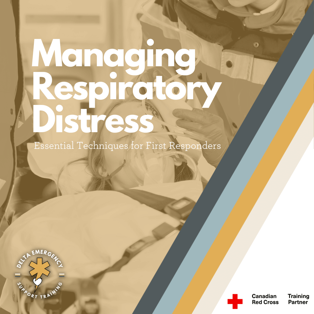
Managing Respiratory Distress: Essential Techniques for First Responders

First responders play a critical role in emergency situations, including managing the breathing of patients. In this blog, we will discuss the importance of breathing management for first responders and provide tips for ensuring the best possible outcomes for patients.

As a first responder, proper breathing management is essential for providing effective care to patients in respiratory distress. Understanding the anatomy of the respiratory system, recognizing signs and symptoms of respiratory distress, and maintaining normal breathing rates are all important aspects of breathing management. In this post, we'll explore these topics in the context of first responders.

The first responder has placed an oxygen mask, known as a non-rebreather, over the patient's nose and mouth. The non-rebreather has a bag that fills with oxygen, which allows the patient to receive a high concentration of oxygen with each breath. The non-rebreather mask is used to treat patients who are experiencing respiratory distress or who require high levels of oxygen.

Anatomy of the Respiratory System

As a first responder, it's important to have a basic understanding of the anatomy of the respiratory system. The respiratory system consists of several parts that work together to facilitate breathing. The main organs involved in breathing are the lungs, which are located in the chest. The lungs are made up of millions of tiny air sacs called alveoli, which are responsible for exchanging gases with the blood. The trachea, or windpipe, connects the lungs to the mouth and nose, while the diaphragm is a muscle that helps with breathing by contracting and relaxing. The bronchi are the tubes that branch off from the trachea and lead to the lungs, and the bronchioles are smaller tubes that branch off from the bronchi and lead to the alveoli.

Signs and Symptoms of Respiratory Distress

As a first responder, it's important to recognize the signs and symptoms of respiratory distress. These may include:

  1. Shortness of breath:

    Feeling like you can't get enough air, or struggling to breathe, is a common symptom of respiratory distress.

  2. Rapid breathing:

    Breathing faster than normal, or taking shallow breaths, may be a sign of respiratory distress.

  3. Chest pain:

    Pain or discomfort in the chest can be a sign of a serious respiratory problem.

  4. Wheezing/Stridor:

    A high-pitched whistling sound when breathing may indicate narrowing of the airways.

  5. Cyanosis:

    This can indicate a lack of oxygen in the blood.

  6. Confusion:

    Lack of oxygen can cause confusion or disorientation.

  7. Fatigue:

    Difficulty breathing can cause fatigue or weakness.

Maintaining Normal Breathing Rates

As a first responder, it's important to know the normal breathing rates for adults and children. The normal breathing rate for an adult is around 12-20 breaths per minute, while the normal breathing rate for a child is higher, around 20-40 breaths per minute. However, certain conditions can cause abnormal breathing rates. For example, respiratory distress can cause rapid, shallow breathing, while other conditions, such as sleep apnea, can cause periods of slowed or paused breathing.

Breathing Management

Proper breathing management is essential for providing effective care to patients in respiratory distress. As a first responder, here are some tips for managing breathing:

  1. Administer oxygen:

    Depending on the severity of the patient's respiratory distress, administering oxygen may be necessary. The appropriate oxygen delivery device should be used based on the patient's level of respiratory distress.

  2. Position the patient:

    Positioning the patient in a way that maximizes their breathing capacity can be helpful. For example, placing the patient in a sitting position with their head slightly elevated may make it easier for them to breathe.

  3. Provide assisted ventilation:

    In some cases, conscious bagging with a bag valve mask may be necessary to help the patient normalize their breathing rate.

  4. Transport the patient to a medical facility:

    If the patient's respiratory distress is severe, transport them to a medical facility for further treatment.

Oxygen masks and flow rates

Breathing masks are a crucial piece of equipment for first responders, as they allow them to provide oxygen to patients in respiratory distress. There are several types of breathing masks available, each with its own specific use and oxygen flow rate. In this post, we'll explore the different types of breathing masks commonly used by first responders, including the nasal cannula, simple face mask, non-rebreather mask, and bag valve mask.

Nasal Cannula

The nasal cannula is a type of oxygen delivery device that is used to provide oxygen to patients with mild to moderate respiratory distress. It is a thin tube that is inserted into the nostrils and is held in place by a strap that goes around the patient's head. The nasal cannula is typically used to deliver low to medium oxygen flow rates, typically ranging from 1 to 6 liters per minute (LPM).

Simple Face Mask

The simple face mask is another type of oxygen delivery device that is used to provide oxygen to patients with respiratory distress. It covers the nose and mouth and is held in place by an elastic strap that goes around the patient's head. The simple face mask is typically used to deliver medium to high oxygen flow rates, typically ranging from 5 to 10 LPM.

Non-Rebreather Mask

The non-rebreather mask is a type of face mask that is used to deliver high concentrations of oxygen to patients with severe respiratory distress. It covers both the nose and mouth and has a one-way valve that allows the patient to inhale oxygen from a reservoir bag. The non-rebreather mask is typically used to deliver high oxygen flow rates, ranging from 10 to 15 LPM.

Bag Valve Mask

The bag valve mask, also known as a manual resuscitator, is a handheld device that is used to provide positive pressure ventilation to patients in respiratory distress. It consists of a bag that is attached to a mask and is used to deliver oxygen to the patient's lungs. The bag valve mask is typically used to deliver high oxygen flow rates, ranging from 10 to 15 LPM.

By following proper breathing management techniques, first responders can help ensure that patients receive the oxygen they need to survive, while also ensuring their own safety in emergency situations.

At Delta Emergency Support Training, we understand the importance of being prepared for medical emergencies. We offer a range of training sessions to help individuals and classes develop the skills and knowledge they need to respond to emergency situations. Our courses include Standard First Aid (SFA), Advanced First Aid (AFA), and Emergency Medical Responder (EMR), and we offer in-person, hybrid, and online options to suit different needs.

If you have any questions about our training sessions or medical emergencies in general, please don't hesitate to reach out to us. You can contact us at info@deltaemergency.com and we'll be happy to assist you.

Read More

Firefighter Training: OPA's and NPA's for Airway Management

Nasopharyngeal airways (NPA) and oropharyngeal airways (OPA) are commonly used in healthcare settings to maintain a patient's airway and assist with breathing.When used correctly, NPAs can help clients who may be experiencing respiratory distress during certain treatments. However, it's important to have the necessary knowledge and protocols in place to safely insert and monitor the device. At Delta Emergency Support Training, we can help you learn how to safely use NPAs in spa settings and beyond. Our training sessions include a range of courses, including Standard First Aid (SFA), Advanced First Aid (AFA), and Emergency Medical Responder (EMR), and we offer in-person, hybrid, and online options to suit different needs. Contact us at info@deltaemergency.com to learn more about our training sessions and how we can help you develop the skills and knowledge you need to respond to emergency situations effectively.

As a firefighter, you need to be prepared to respond to emergency situations, including those involving airway management for an unconscious patients airway. Two commonly used tools for airway management are oropharyngeal airways (OPA's) and nasopharyngeal airways (NPA's). Today we'll discuss what these devices are, why they are used, and how to properly insert them as a first responder.

Four colour coded Oropharyngeal airways lay across a medical sheet.

What are OPA's and NPA's?

Oropharyngeal airways (OPA's) and nasopharyngeal airways (NPA's) are medical devices used to keep an airway open in patients who can’t secure it themselves. They are used when the patient is unable to maintain their own airway due to injury, illness, or drug overdose and become unconscious. OPA's and NPA's are both designed to be inserted into the patient's airway to help keep it open.

An OPA is a curved plastic device that is inserted into the patient's mouth and extends into the pharynx. It helps to prevent the tongue from obstructing the airway and allows air to pass freely into the lungs. OPA's are available in different sizes to accommodate patients of different ages and sizes.

An NPA is a flexible, hollow tube that is inserted into the patient's nostril and extends into the nasopharynx. It helps to keep the airway open and can be used in patients who are unable to tolerate an OPA due to gag reflex or other reasons.

Why do we use OPA's and NPA's?

The primary reason for using OPA's and NPA's is to maintain a patient's airway and ensure that they are able to breathe. When a patient's airway is obstructed, it can quickly lead to hypoxia, which is a dangerous condition where the body's tissues do not receive enough oxygen. OPA's and NPA's are used to prevent this from happening by keeping the airway open and allowing oxygen to flow freely into the lungs.

Testing for LOC (Level Of Consciousness)

Before inserting an OPA, it is important to assess the patient's level of consciousness. The AVPU scale is a simple and quick method for assessing a patient's level of consciousness based on their response to stimuli. The scale has four categories:

  • A: Alert

  • V: Verbal response

  • P: Pain response

  • U: Unresponsive

To use the AVPU scale, a healthcare provider would first try to get the patient's attention by speaking to them in a normal tone of voice. If the patient responds appropriately, they would be classified as "A" for alert. If the patient responds to verbal stimuli but is not fully alert, they would be classified as "V" for verbal response. If the patient only responds to painful/physical stimuli, such as a trap squeeze or nail bed pressure, they would be classified as "P" for pain/physical response. If the patient does not respond to any stimuli, they would be classified as "U" for unresponsive.

If a patient is assessed as being unresponsive or only responding to painful stimuli, an OPA may need to be inserted to maintain their airway. However, if the patient is alert or responding to verbal stimuli, an OPA may not be necessary.

How to insert OPA's and NPA's as a first responder

Proper insertion of OPA's and NPA's is crucial to their effectiveness and patient safety. Here are some general steps for inserting OPA's and NPA's:

Inserting an OPA:

  1. Choose the appropriate size OPA for the patient based on their age and size. Measure from the corner of the mouth to the angle of the jaw.

  2. Position the patient's head in a neutral position.

  3. Open the patient's mouth using the cross finger technique.

  4. Insert the OPA into the patient's mouth, with the curved end facing the roof of the mouth.

  5. Gently rotate the device 180 degrees as you insert it, until it is in a anatomical position.

Inserting an NPA:

  1. Choose the appropriate size NPA for the patient based on their age and size by measuring the tip of the nose to the corner of the jaw.

  2. Lubricate the NPA with a water-soluble lubricant.

  3. Position the patient's head in a neutral position.

  4. Insert the NPA into the patient's nostril starting with the right nostril. Bevelled edge facing the septum.

  5. Gently advance the device until it reaches the back of the nasopharynx

At Delta Emergency Support Training, we understand the importance of being prepared for medical emergencies. We offer a range of training sessions to help individuals and classes develop the skills and knowledge they need to respond to emergency situations. Our courses include Standard First Aid (SFA), Advanced First Aid (AFA), and Emergency Medical Responder (EMR), and we offer in-person, hybrid, and online options to suit different needs.

If you have any questions about our training sessions or medical emergencies in general, please don't hesitate to reach out to us. You can contact us at info@deltaemergency.com and we'll be happy to assist you.

Read More

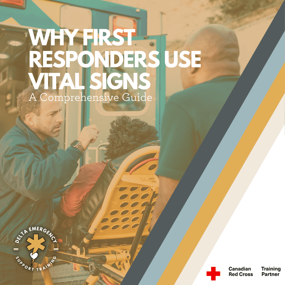
Why First Responders Use Vital Signs: A comprehensive guide

Vital signs are measurements of the body's basic functions and are used to assess a person's overall health and wellbeing. There are four primary vital signs: body temperature, pulse rate, blood pressure, and respiratory rate. Each vital sign provides important information about a person's health and can help identify underlying health conditions or other problems.

Body temperature is a measure of the body's internal heat and is typically measured using a thermometer. A fever is generally defined as a body temperature above 100.4°F (38°C) and can indicate an infection or other underlying health condition.

Pulse rate is a measure of the heart rate, or the number of times the heart beats per minute. A rapid pulse may indicate a fever, dehydration, or an irregular heartbeat, while a slow pulse may indicate heart disease or other health conditions.

Blood pressure is a measure of the force of blood against the walls of arteries as the heart pumps blood through the body. High blood pressure, or hypertension, can increase the risk of heart disease, stroke, and other health problems.

Respiratory rate is a measure of the number of breaths a person takes per minute. A rapid respiratory rate may indicate an underlying health condition, such as asthma or pneumonia.

Monitoring vital signs is an essential part of healthcare and can help healthcare professionals and first responders make informed decisions about treatment and care.

Vital signs are measurements of basic bodily functions that are essential for assessing a person's overall health and well-being. These measurements are typically taken by healthcare professionals, but they can also be taken by trained first responders in emergency situations.

first responders attending to a patient lying on a cot outside an ambulance. One responder is checking the patient's vital signs while the other is preparing medical equipment. The patient appears to be receiving urgent medical attention, and the responders are focused and prepared to administer necessary treatment

What Are Vital Signs?

Vital signs are a set of measurements that healthcare professionals use to assess a patient's overall health status. These measurements include temperature, heart rate, blood pressure, respiratory rate, Glasgow Coma Scale (GCS), oxygen saturation (SPO2), pupils and skin.

  1. Body Temperature:

    Body temperature is an important vital sign as it can indicate whether a person is healthy or if they have a fever, which could be a sign of infection. Body temperature can be measured in several ways, including orally, rectally, or using a forehead thermometer. It is important to use the correct method and follow the manufacturer's instructions when taking a person's temperature.

  2. Heart Rate:

    Heart rate: Heart rate is the number of times the heart beats per minute and is a reflection of the heart's ability to circulate blood throughout the body. The heart rate can vary depending on age, gender, physical activity, and overall health status. A high heart rate could indicate a potential problem, such as an irregular heartbeat, dehydration, or anxiety, while a low heart rate could be a sign of a heart condition or medication side effect.

  3. Blood Pressure:

    Blood pressure is the measure of the force of blood against the walls of the arteries. It is measured in millimetres of mercury (mmHg) and consists of two numbers: the systolic pressure (the higher number) and the diastolic pressure (the lower number). A healthy blood pressure reading is typically around 120/80 mmHg. High blood pressure (hypertension) can put a strain on the heart and blood vessels, while low blood pressure (hypotension) can cause dizziness, fainting, or other symptoms.

  4. Respiratory Rate:

    Respiratory rate is the number of breaths taken per minute and is a reflection of the body's ability to take in oxygen and expel carbon dioxide. The respiratory rate can vary depending on age, physical activity, and overall health status. A high respiratory rate could indicate a potential problem, such as a lung infection or asthma, while a low respiratory rate could be a sign of respiratory depression or a medical condition that affects breathing.

  5. Glasgow Coma Scale:

    Glasgow Coma Scale or GCS measures a patient's response to verbal, motor, and eye-opening stimuli. The score ranges from 3 to 15, with a higher score indicating a higher level of consciousness. The GCS is commonly used in trauma settings to assess patients with brain injury, altered mental status, or who are sedated.

  6. SPO2:

    SPO2 is a measure of the amount of oxygen that is carried by the hemoglobin in the red blood cells. The measurement of SPO2 is non-invasive and can be performed using a pulse oximeter, which is a small device that is attached to the finger, toe, or earlobe. The pulse oximeter uses light to measure the amount of oxygen that is present in the blood.

  7. Pupils:

    Pupils can also be an important vital sign in assessing a person's neurological function. The size, shape, and reactivity of the pupils can provide valuable information about the health of the brain and nervous system. Unequal pupil size (ani or a lack of reactivity to light can be a sign of a neurological issue, such as a brain injury or nerve damage.

  8. Skin:

    Assessing skin color and moisture is important because changes in these vital signs can be indicative of underlying medical conditions. For example, changes in skin color can be a sign of poor blood flow, infection, or inflammation, while changes in skin moisture can be a sign of dehydration or certain medical conditions.

Normal Vital Sign Ranges

The normal ranges for vital signs vary depending on a person's age, gender, and health status. Here are the standard ranges:

  1. Body Temperature:

    A normal body temperature generally ranges from 36.1°C to 37.2°C, with an average of 37°C. A fever, which is typically defined as a body temperature above 38°C, can be a sign of an infection or illness. Hypothermia, in which the body's core temperature drops below 35°C, can be caused by prolonged exposure to cold temperatures or certain medical conditions. Hyperthermia, in which the body's core temperature rises above its normal range, typically above 40°C, can be caused by heatstroke, extreme physical exertion, or certain medications.

  2. Pulse Rate:

    The normal resting heart rate range for an adult is between 60 to 100 beats per minute. A higher or lower heart rate may indicate a medical condition. Healthcare professionals use heart rate to assess cardiac function, diagnose heart conditions, and monitor the effectiveness of treatments.

  3. Blood Pressure:

    The normal blood pressure for an adult is typically around 120/80 mmHg. High blood pressure, or hypertension, is defined as consistently measuring higher than 130/80 mmHg, and can increase the risk of heart disease, stroke, and other health problems. Low blood pressure, or hypotension, is less common but can also be a serious medical condition, and is typically defined as a systolic pressure of less than 90 mmHg or a diastolic pressure of less than 60 mmHg.

  4. Respiratory Rate:

    The normal respiratory rate for an adult at rest is typically between 12 and 20 breaths per minute. A higher or lower respiratory rate than this range may indicate a medical condition. The quality of breath is also important in assessing a patient's respiratory function. Shallow, labored breathing or irregular breathing patterns can indicate respiratory distress and may require immediate medical attention.

  5. Glasgow Coma Scale:

    A GCS score of 15 is considered normal, indicating that the person is fully alert and oriented. The GCS is based on three categories: eye opening, verbal response, and motor response, and each category is assigned a score from 1-4 or 1-6. A higher score indicates a higher level of consciousness, while a lower score indicates a lower level of consciousness.

  6. SPO2

    Healthy SPO2 levels are between 95% and 100%, and SPO2 levels below 90% can indicate a lack of oxygen in the body. Supplemental oxygen may be necessary to raise the patient's SPO2 levels to a healthy range, but it is important to monitor the oxygen levels carefully and avoid over-oxygenation. Healthcare professionals use SPO2 measurements to guide treatment decisions and ensure that patients are receiving the appropriate level of oxygen.

  7. Pupils:

    The normal range for pupil size is between 2-4 millimeters in diameter for both pupils, and they should be equal in size. Pupils that are larger or smaller than this range, or are unequal in size (anisocoria), can be indicative of underlying medical conditions. Additionally, pupils should react briskly to changes in light, which is known as the pupillary light reflex. A sluggish or absent pupillary light reflex can be a sign of a neurological issue, such as a brain injury or nerve damage.

  8. Skin:

    Skin can vary in color and moisture depending on a person's age, sex, ethnicity, and overall health. In general, healthy skin should have a consistent color and texture across the body, without any unusual bumps, rashes, or lesions. The normal range of skin moisture varies from person to person, but it should generally feel soft and supple, without feeling excessively dry or oily.

Why First Responders Use Vital Signs?

Interpreting vital signs can help first responders make informed decisions about the best course of action for treating a patient. Monitoring changes in vital signs over time can also help to identify trends or progression of medical conditions, and inform treatment plans for better outcomes. First responders are trained to assess and respond to changes in vital signs quickly and effectively, which is essential in emergency situations.

At Delta Emergency Support Training we are happy to answer any questions you may have about vital signs as well as training sessions for individuals or classes. We offer in person, hybrid and online classes for Standard First Aid (SFA) Advanced First Aid (AFA) and Emergency Medical Responder (EMR) courses. For all inquiries please email info@deltaemergency.com .

Read More