The Delta Dispatch

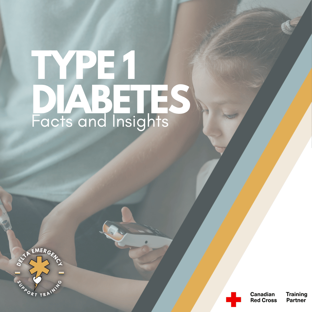
Type 1 Diabetes: Insights into its Origins, Signs, and Challenges

Type 1 diabetes, where the pancreas's struggle with insulin production takes center stage. This blog peels back the layers to reveal the mysterious causes behind this chronic condition, providing valuable insights into recognizing symptoms early. As we navigate the complications that can arise if left unchecked, we also explore the ongoing research endeavors aiming to usher in a new era of preventive strategies. Join us in decoding the intricacies of Type 1 diabetes, empowering individuals and their families with the knowledge needed to navigate this journey with resilience and hope.

Introduction:

LITTLE GIRL HAVING BLOOD SUGAR CHECKED BY HER MOTHER.

In our exploration of Type 1 diabetes, previously recognized as juvenile diabetes or insulin-dependent diabetes, we uncover a complex chronic condition where the pancreas produces insufficient insulin. Insulin, a key player in energy production, allows glucose to enter cells. This blog aims to delve into the intricacies of Type 1 diabetes, examining its causes, symptoms, complications, risk factors, and the ongoing quest for preventive strategies.

Unraveling Causes and Risk Factors:

The mystery behind the onset of Type 1 diabetes persists, with researchers pointing towards a potential glitch in the immune system. Rather than safeguarding the body, the immune system turns on the insulin-producing cells in the pancreas. Genetic factors, exposure to certain viruses, and environmental elements may contribute to the development of Type 1 diabetes. Risk factors include a familial history of the condition, specific genetic predispositions, geographical factors, and age, with noticeable peaks in children aged 4-7 and 10-14.

Insulin and Glucose Symphony:

The pivotal hormone, insulin, originating from the pancreas, orchestrates the delicate balance of blood sugar levels. In the absence of insulin, as seen in Type 1 diabetes, glucose remains stranded outside cells, leading to elevated blood sugar levels. Glucose, sourced from food and the liver, serves as the primary energy provider for cells. In Type 1 diabetes, the dearth of insulin results in the buildup of sugar in the bloodstream, presenting a potential hazard of life-threatening complications.

Recognizing Symptoms and Diagnosis:

Type 1 diabetes is characterized by abrupt symptoms such as increased thirst, frequent urination, unexpected weight loss, irritability, and blurry vision. Seeking prompt medical attention is crucial if these symptoms manifest. Diagnosis involves thorough blood tests to measure blood sugar levels and assess other critical parameters.

Unveiling Complications:

Untamed Type 1 diabetes can lead to severe complications impacting vital organs:

  1. Heart and Blood Vessel Disease: Elevates the risk of coronary artery disease, heart attack, stroke, atherosclerosis, and high blood pressure.

  2. Nerve Damage (Neuropathy): Can induce tingling, numbness, burning, or pain, particularly in the extremities. Digestive system nerves' damage may result in gastrointestinal issues.

  3. Kidney Damage (Nephropathy): Poses a risk of kidney failure or end-stage kidney disease.

  4. Eye Damage: May result in diabetic retinopathy, cataracts, glaucoma, and potential blindness.

  5. Foot Damage: Increases the risk of severe foot complications, potentially leading to amputation if untreated.

  6. Skin and Mouth Conditions: Enhances susceptibility to skin and mouth infections, including bacterial and fungal infections, gum disease, and dry mouth.

  7. Pregnancy Complications: Poorly controlled diabetes during pregnancy heightens the risk of various complications.

Preventive Measures and Management:

Currently, no foolproof method exists to prevent Type 1 diabetes. Nevertheless, ongoing research endeavors to thwart the disease or mitigate islet cell damage in newly diagnosed individuals. Clinical trials exploring preventive measures offer hope, and individuals diagnosed with Type 1 diabetes should discuss potential participation with their healthcare providers.

Conclusion:

Type 1 diabetes poses a unique set of challenges necessitating lifelong management. By comprehending its origins, symptoms, complications, and ongoing research initiatives, individuals and their families can navigate this journey with resilience. Collaboration between healthcare providers and individuals is crucial for effective management, and as research progresses, optimism grows for advancements in treatment and prevention strategies.

Read More

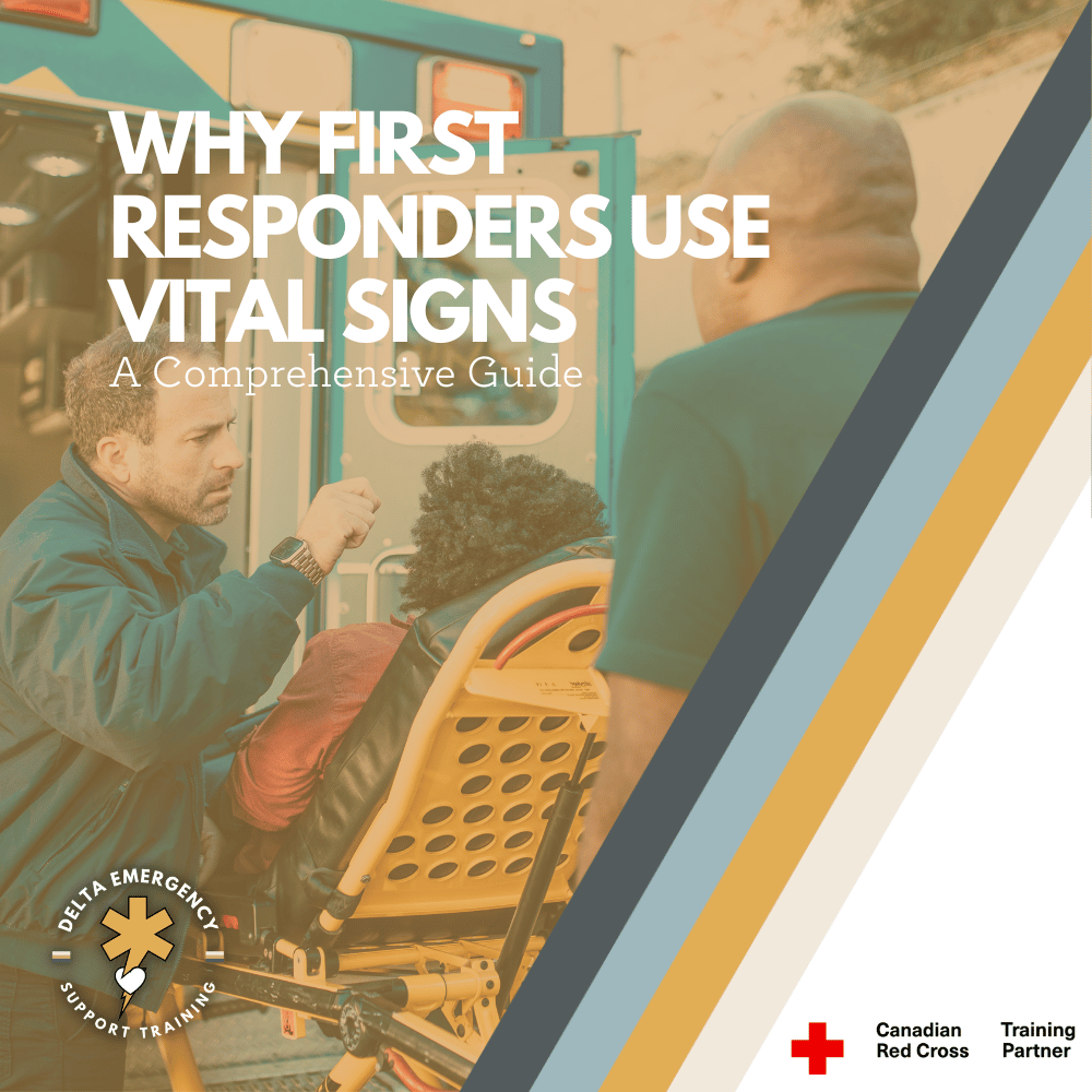
Why First Responders Use Vital Signs: A comprehensive guide

Vital signs are measurements of the body's basic functions and are used to assess a person's overall health and wellbeing. There are four primary vital signs: body temperature, pulse rate, blood pressure, and respiratory rate. Each vital sign provides important information about a person's health and can help identify underlying health conditions or other problems.

Body temperature is a measure of the body's internal heat and is typically measured using a thermometer. A fever is generally defined as a body temperature above 100.4°F (38°C) and can indicate an infection or other underlying health condition.

Pulse rate is a measure of the heart rate, or the number of times the heart beats per minute. A rapid pulse may indicate a fever, dehydration, or an irregular heartbeat, while a slow pulse may indicate heart disease or other health conditions.

Blood pressure is a measure of the force of blood against the walls of arteries as the heart pumps blood through the body. High blood pressure, or hypertension, can increase the risk of heart disease, stroke, and other health problems.

Respiratory rate is a measure of the number of breaths a person takes per minute. A rapid respiratory rate may indicate an underlying health condition, such as asthma or pneumonia.

Monitoring vital signs is an essential part of healthcare and can help healthcare professionals and first responders make informed decisions about treatment and care.

Vital signs are measurements of basic bodily functions that are essential for assessing a person's overall health and well-being. These measurements are typically taken by healthcare professionals, but they can also be taken by trained first responders in emergency situations.

first responders attending to a patient lying on a cot outside an ambulance. One responder is checking the patient's vital signs while the other is preparing medical equipment. The patient appears to be receiving urgent medical attention, and the responders are focused and prepared to administer necessary treatment

What Are Vital Signs?

Vital signs are a set of measurements that healthcare professionals use to assess a patient's overall health status. These measurements include temperature, heart rate, blood pressure, respiratory rate, Glasgow Coma Scale (GCS), oxygen saturation (SPO2), pupils and skin.

  1. Body Temperature:

    Body temperature is an important vital sign as it can indicate whether a person is healthy or if they have a fever, which could be a sign of infection. Body temperature can be measured in several ways, including orally, rectally, or using a forehead thermometer. It is important to use the correct method and follow the manufacturer's instructions when taking a person's temperature.

  2. Heart Rate:

    Heart rate: Heart rate is the number of times the heart beats per minute and is a reflection of the heart's ability to circulate blood throughout the body. The heart rate can vary depending on age, gender, physical activity, and overall health status. A high heart rate could indicate a potential problem, such as an irregular heartbeat, dehydration, or anxiety, while a low heart rate could be a sign of a heart condition or medication side effect.

  3. Blood Pressure:

    Blood pressure is the measure of the force of blood against the walls of the arteries. It is measured in millimetres of mercury (mmHg) and consists of two numbers: the systolic pressure (the higher number) and the diastolic pressure (the lower number). A healthy blood pressure reading is typically around 120/80 mmHg. High blood pressure (hypertension) can put a strain on the heart and blood vessels, while low blood pressure (hypotension) can cause dizziness, fainting, or other symptoms.

  4. Respiratory Rate:

    Respiratory rate is the number of breaths taken per minute and is a reflection of the body's ability to take in oxygen and expel carbon dioxide. The respiratory rate can vary depending on age, physical activity, and overall health status. A high respiratory rate could indicate a potential problem, such as a lung infection or asthma, while a low respiratory rate could be a sign of respiratory depression or a medical condition that affects breathing.

  5. Glasgow Coma Scale:

    Glasgow Coma Scale or GCS measures a patient's response to verbal, motor, and eye-opening stimuli. The score ranges from 3 to 15, with a higher score indicating a higher level of consciousness. The GCS is commonly used in trauma settings to assess patients with brain injury, altered mental status, or who are sedated.

  6. SPO2:

    SPO2 is a measure of the amount of oxygen that is carried by the hemoglobin in the red blood cells. The measurement of SPO2 is non-invasive and can be performed using a pulse oximeter, which is a small device that is attached to the finger, toe, or earlobe. The pulse oximeter uses light to measure the amount of oxygen that is present in the blood.

  7. Pupils:

    Pupils can also be an important vital sign in assessing a person's neurological function. The size, shape, and reactivity of the pupils can provide valuable information about the health of the brain and nervous system. Unequal pupil size (ani or a lack of reactivity to light can be a sign of a neurological issue, such as a brain injury or nerve damage.

  8. Skin:

    Assessing skin color and moisture is important because changes in these vital signs can be indicative of underlying medical conditions. For example, changes in skin color can be a sign of poor blood flow, infection, or inflammation, while changes in skin moisture can be a sign of dehydration or certain medical conditions.

Normal Vital Sign Ranges

The normal ranges for vital signs vary depending on a person's age, gender, and health status. Here are the standard ranges:

  1. Body Temperature:

    A normal body temperature generally ranges from 36.1°C to 37.2°C, with an average of 37°C. A fever, which is typically defined as a body temperature above 38°C, can be a sign of an infection or illness. Hypothermia, in which the body's core temperature drops below 35°C, can be caused by prolonged exposure to cold temperatures or certain medical conditions. Hyperthermia, in which the body's core temperature rises above its normal range, typically above 40°C, can be caused by heatstroke, extreme physical exertion, or certain medications.

  2. Pulse Rate:

    The normal resting heart rate range for an adult is between 60 to 100 beats per minute. A higher or lower heart rate may indicate a medical condition. Healthcare professionals use heart rate to assess cardiac function, diagnose heart conditions, and monitor the effectiveness of treatments.

  3. Blood Pressure:

    The normal blood pressure for an adult is typically around 120/80 mmHg. High blood pressure, or hypertension, is defined as consistently measuring higher than 130/80 mmHg, and can increase the risk of heart disease, stroke, and other health problems. Low blood pressure, or hypotension, is less common but can also be a serious medical condition, and is typically defined as a systolic pressure of less than 90 mmHg or a diastolic pressure of less than 60 mmHg.

  4. Respiratory Rate:

    The normal respiratory rate for an adult at rest is typically between 12 and 20 breaths per minute. A higher or lower respiratory rate than this range may indicate a medical condition. The quality of breath is also important in assessing a patient's respiratory function. Shallow, labored breathing or irregular breathing patterns can indicate respiratory distress and may require immediate medical attention.

  5. Glasgow Coma Scale:

    A GCS score of 15 is considered normal, indicating that the person is fully alert and oriented. The GCS is based on three categories: eye opening, verbal response, and motor response, and each category is assigned a score from 1-4 or 1-6. A higher score indicates a higher level of consciousness, while a lower score indicates a lower level of consciousness.

  6. SPO2

    Healthy SPO2 levels are between 95% and 100%, and SPO2 levels below 90% can indicate a lack of oxygen in the body. Supplemental oxygen may be necessary to raise the patient's SPO2 levels to a healthy range, but it is important to monitor the oxygen levels carefully and avoid over-oxygenation. Healthcare professionals use SPO2 measurements to guide treatment decisions and ensure that patients are receiving the appropriate level of oxygen.

  7. Pupils:

    The normal range for pupil size is between 2-4 millimeters in diameter for both pupils, and they should be equal in size. Pupils that are larger or smaller than this range, or are unequal in size (anisocoria), can be indicative of underlying medical conditions. Additionally, pupils should react briskly to changes in light, which is known as the pupillary light reflex. A sluggish or absent pupillary light reflex can be a sign of a neurological issue, such as a brain injury or nerve damage.

  8. Skin:

    Skin can vary in color and moisture depending on a person's age, sex, ethnicity, and overall health. In general, healthy skin should have a consistent color and texture across the body, without any unusual bumps, rashes, or lesions. The normal range of skin moisture varies from person to person, but it should generally feel soft and supple, without feeling excessively dry or oily.

Why First Responders Use Vital Signs?

Interpreting vital signs can help first responders make informed decisions about the best course of action for treating a patient. Monitoring changes in vital signs over time can also help to identify trends or progression of medical conditions, and inform treatment plans for better outcomes. First responders are trained to assess and respond to changes in vital signs quickly and effectively, which is essential in emergency situations.

At Delta Emergency Support Training we are happy to answer any questions you may have about vital signs as well as training sessions for individuals or classes. We offer in person, hybrid and online classes for Standard First Aid (SFA) Advanced First Aid (AFA) and Emergency Medical Responder (EMR) courses. For all inquiries please email info@deltaemergency.com .

Read More

Unraveling the Mystery of the Heart's Electrical Dance: How Your Heart Beats to Its Own Rhythm!

Heart's Electrical Conduction - Key Concepts and Terminology. Delve into the intricacies of the heart's electrical conduction system with our comprehensive overview. Learn about the physiology, function, and regulation of the heart's electrical pathways. Gain a deeper understanding of this vital organ's complex conduction system with our informative blog.

Understanding the Intricacies of the Heart's Electrical Conduction System: A Comprehensive Overview

The human heart is a complex organ responsible for pumping blood throughout the body to sustain life. At the core of this remarkable organ lies a sophisticated electrical conduction system that coordinates the heartbeat and ensures its proper functioning. This paper aims to provide a comprehensive overview of the heart's electrical conduction system, including its anatomy, physiology, and the sequence of events that occur during each cardiac cycle.

Anatomy of the Heart's Electrical Conduction System: The heart's electrical conduction system comprises specialized cardiac cells that are distributed in various regions of the heart. These cells possess unique properties that enable them to generate, conduct, and regulate electrical signals, ultimately controlling the rhythm and rate of the heartbeat.

  1. Sinoatrial Node (SA Node): The SA node is located in the upper right atrium near the opening of the superior vena cava. Often referred to as the "natural pacemaker" of the heart, the SA node initiates the electrical impulses that determine the heart's rhythm. It generates electrical signals that travel through the atria, causing them to contract and pump blood into the ventricles.

  2. Atrioventricular Node (AV Node): The AV node is located at the bottom of the right atrium near the interatrial septum. It acts as a gatekeeper, delaying the electrical signals from the atria before passing them to the ventricles. This delay allows the atria to fully contract and empty their blood into the ventricles before the ventricles contract.

  3. Bundle of His: The Bundle of His is a collection of specialized fibers that transmit the electrical signals from the AV node to the ventricles. It branches into the left and right bundle branches, which extend along the interventricular septum and eventually divide into smaller Purkinje fibers.

  4. Purkinje Fibers: Purkinje fibers are specialized cardiac cells that distribute the electrical signals throughout the ventricles, causing them to contract and pump blood out of the heart.

Physiology of the Heart's Electrical Conduction System: The electrical activity in the heart is regulated by the flow of ions across the cell membranes of the cardiac cells, which results in changes in the cell's electrical charge. These changes in electrical charge create the electrical signals that propagate through the heart and coordinate the heartbeat.

  1. Resting Membrane Potential: The resting membrane potential of cardiac cells is around -90 millivolts (mV), meaning that the inside of the cell is more negatively charged compared to the outside. This is due to the uneven distribution of ions across the cell membrane, with higher concentrations of potassium (K+) inside the cell and higher concentrations of sodium (Na+) and calcium (Ca2+) outside the cell.

  2. Depolarization: The depolarization of cardiac cells is the process by which the cell's electrical charge becomes more positive, leading to the generation of an action potential. The depolarization of the SA node is initiated by the influx of calcium ions, which triggers the opening of voltage-gated sodium channels, allowing sodium ions to rush into the cell. This leads to a rapid depolarization of the cell membrane, resulting in the generation of an action potential.

  3. Action Potential Propagation: Once the action potential is generated in the SA node, it spreads through the atria, causing them to contract. The action potential then reaches the AV node, where it is delayed for a brief period to allow the atria to fully contract and empty their blood into the ventricles. After the delay, the action potential is transmitted through the Bundle of His and the Purkinje fibers, which rapidly conduct the electrical signals to the ventricles. The depolarization of the ventricles leads to their contraction and the ejection of blood from the heart.

  4. Repolarization: After depolarization, the cardiac cells need to repolarize to restore their resting membrane potential and prepare for the next electrical signal. Repolarization is achieved through the movement of potassium ions out of the cell, which results in the restoration of the negative charge inside the cell.

Sequence of Events during Cardiac Cycle:

The electrical conduction system of the heart plays a crucial role in coordinating the events of the cardiac cycle, which consists of two main phases: diastole and systole.

  1. Diastole: During diastole, the heart is relaxed, and the chambers fill with blood. The electrical signals generated by the SA node initiate the diastole by causing the atria to contract and pump blood into the ventricles. The electrical signals then reach the AV node, where there is a slight delay before the signals are transmitted to the ventricles through the Bundle of His and Purkinje fibers. This delay allows for complete filling of the ventricles before their contraction.

  2. Systole: During systole, the heart contracts, and blood is ejected from the ventricles into the arteries. The electrical signals from the Purkinje fibers cause the ventricles to contract and pump blood out of the heart. Once the electrical signals are completed, the heart returns to diastole, and the cycle restarts.

Regulation of the Heart's Electrical Conduction System: The heart's electrical conduction system is regulated by various factors to maintain the proper rhythm and rate of the heartbeat.

  1. Autonomic Nervous System: The autonomic nervous system, consisting of the sympathetic and parasympathetic divisions, plays a significant role in regulating the heart's electrical conduction system. The sympathetic division increases the heart rate and conduction velocity, while the parasympathetic division decreases the heart rate and conduction velocity.

  2. Hormones: Hormones such as adrenaline and noradrenaline released during times of stress or excitement can affect the heart's electrical conduction system, leading to an increase in heart rate and conduction velocity.

  3. Electrolyte Balance: Proper electrolyte balance, particularly the levels of potassium, sodium, and calcium, is crucial for the normal functioning of the heart's electrical conduction system. Disturbances in electrolyte levels can disrupt the conduction of electrical signals, leading to arrhythmias.

The heart's electrical conduction system is a complex and intricate system that ensures the coordinated contraction and relaxation of the heart, leading to effective pumping of blood. The SA node acts as the natural pacemaker, generating electrical signals that initiate and regulate the heartbeat. The electrical signals are conducted through the AV node, Bundle of His, and Purkinje fibers, causing the atria and ventricles to contract in a synchronized manner. Proper regulation of the heart's electrical conduction system is essential for maintaining a healthy heart rhythm and preventing arrhythmias. Further research and understanding of this complex system may lead to advancements in diagnosing and treating various cardiac conditions related to electrical conduction abnormalities.

Read More

Saving Lives: The Vital Role of Automated External Defibrillators (AEDs) in Cardiac Emergency Response

The importance of Automated External Defibrillators (AEDs) in cardiac emergency response cannot be overstated. This powerful photo depicts a person using an AED to deliver life-saving defibrillation during a critical moment of a cardiac emergency. With a descriptive filename, alt text, and metadata that includes relevant keywords, this image serves as a powerful visual representation of the vital role of AEDs in saving lives during cardiac emergencies.

Saving Lives with Automated External Defibrillators (AEDs): What You Need to Know

The importance of Automated External Defibrillators (AEDs) in cardiac emergency response cannot be overstated. This photo depicts an AED, ready to deliver life-saving defibrillation during a critical moment of a cardiac emergency.

Cardiac arrest is a sudden and life-threatening condition that occurs when the heart suddenly stops beating effectively. Immediate intervention is critical to increase the chances of survival, and Automated External Defibrillators (AEDs) have emerged as a crucial tool in saving lives during cardiac emergencies. In this blog post, we will explore what AEDs are, how they work, and why they are essential in cardiac arrest management.

What are AEDs?

An Automated External Defibrillator (AED) is a portable electronic device that is used to diagnose and treat life-threatening cardiac arrhythmias, such as ventricular fibrillation (VF) and pulseless ventricular tachycardia (VT). AEDs are designed to be used by non-medical professionals, including laypersons, and are commonly found in public places, workplaces, airports, schools, and other high-traffic areas.

How do AEDs work?

AEDs work by delivering an electric shock to the heart to restore its normal rhythm. When a person experiences cardiac arrest, the heart's electrical activity becomes chaotic, causing the heart to quiver or fibrillate instead of pumping blood effectively. AEDs are equipped with built-in sensors and computer algorithms that analyze the heart's electrical activity through adhesive pads (called electrodes) placed on the person's chest. If a shockable rhythm, such as Ventricular Fibrillation (VF) or Ventricular Tachycardia (VT), is detected, the AED will prompt the user to deliver a shock by pressing a button or automatically deliver the shock if it is a fully automated AED.

An Automated External Defibrillator (AED) is a portable electronic device that is used to diagnose and treat life-threatening cardiac arrhythmias, such as ventricular fibrillation (VF) and pulseless ventricular tachycardia (VT).

The shock delivered by the AED is intended to stop the chaotic electrical activity in the heart and allow the heart to reset itself and resume a normal rhythm. AEDs are designed to be safe and effective, with automated features that guide the user through each step of the process, including providing voice prompts, visual instructions, and feedback on the quality of CPR (cardiopulmonary resuscitation) being performed.

Why are AEDs important?

AEDs play a crucial role in improving the chances of survival for individuals experiencing sudden cardiac arrest. According to the American Heart Association (AHA), the use of an AED, in combination with CPR, can significantly increase the survival rate of cardiac arrest victims. In fact, for every minute that passes without defibrillation, the chances of survival decrease by approximately 10%. Therefore, quick access to an AED and early defibrillation is critical to increasing the chances of survival.


An Automated External Defibrillator (AED) electricity in action, ready to provide life-saving defibrillation during a cardiac emergency.


They are designed to be used by anyone, regardless of their medical training or experience. They are user-friendly, with clear instructions and prompts that guide the user through the entire process. Many AEDs also have visual aids, such as diagrams or animations, to assist users in correct pad placement and usage. This makes AEDs accessible and empowers bystanders to take immediate action during a cardiac emergency, even before professional medical help arrives.

AEDs are also lightweight and portable, making them easy to transport and use in various settings. They are often designed with durable cases, built-in carrying handles, and sometimes even equipped with backpacks or wall-mounted cabinets for easy storage and accessibility. Many AEDs also perform regular self-checks and have indicators that show their readiness status, ensuring that they are always ready for use in an emergency.

In addition to their ease of use and portability, AEDs are cost-effective. The cost of AEDs has decreased over the years, making them more affordable and accessible for public use. Some AEDs also come with maintenance packages, including


Another important aspect of AEDs is the potential for widespread community impact. When AEDs are strategically placed in public locations, they can be easily accessed and used by bystanders in the critical minutes following a cardiac arrest. This can significantly shorten the time to defibrillation and increase the chances of survival. Many communities, workplaces, and public facilities have recognized the importance of having AEDs readily available and have implemented AED programs as part of their emergency response plans. These AED programs often include training initiatives to educate community members on how to recognize cardiac arrest, perform CPR, and use an AED effectively, further empowering the community to respond to cardiac emergencies.

In conclusion, Automated External Defibrillators (AEDs) are crucial tools in saving lives during cardiac emergencies. Their ease of use, portability, affordability, and potential for widespread community impact make them essential components of cardiac arrest management. By providing early defibrillation and increasing the chances of survival, AEDs empower bystanders to take action and make a significant difference in saving lives. If you encounter someone experiencing sudden cardiac arrest, remember to call emergency services, start CPR, and locate and use an AED as soon as possible for the best chance of survival.

Read More
First Aid Instructor, Professional Responder Jarrett Chisholm First Aid Instructor, Professional Responder Jarrett Chisholm

Unlock Your Potential: Become a Certified First Aid Instructor with Canadian Red Cross

Once you have successfully completed the First Aid Instructor course and obtained certification, you can start teaching Canadian Red Cross first aid and CPR courses. As a First Aid Instructor, you have the opportunity to share life-saving skills with…

Becoming a Canadian Red Cross First Aid Instructor: Training, Support, and Impact

Are you passionate about first aid and CPR education? Do you want to make a meaningful impact in your community? Becoming a Canadian Red Cross First Aid Instructor can provide you with the training, support, and resources to share life-saving skills and contribute to building safer and more resilient communities. In this post, we will explore the various aspects of becoming a Canadian Red Cross First Aid Instructor, including the Teaching Experience Supervisor (TES), Training Partners, Instructor Development Centers (IDCs), and the opportunities for professional development and networking.

Requirements for Becoming a First Aid Instructor

Becoming a Canadian Red Cross First Aid Instructor requires meeting certain prerequisites. To be eligible for instructor certification, you must hold a valid Canadian Red Cross certification in the respective first aid and/or CPR course you wish to teach, such as Standard First Aid, Emergency First Aid, or CPR/AED. You must also have a minimum age requirement of 18 years and demonstrate proficiency in the course content and skills. Additionally, you should possess good communication and teaching skills, as well as a passion for promoting health and safety in your community.

Training Process

The Canadian Red Cross offers comprehensive training programs for aspiring First Aid Instructors. The training process typically involves a series of steps, including completing an instructor application, attending an instructor course, and successfully passing a practical and written exam. The instructor course provides in-depth training on the course content, instructional techniques, adult learning principles, and evaluation methods. It equips you with the necessary knowledge and skills to effectively teach first aid and CPR courses, and prepares you to deliver courses in various settings, such as classrooms, workplaces, and community organizations.

Teaching Opportunities

Once you have successfully completed the First Aid Instructor course and obtained certification, you can start teaching Canadian Red Cross first aid and CPR courses. As a First Aid Instructor, you have the opportunity to share life-saving skills with individuals and groups, empowering them to respond confidently and effectively in emergency situations. You can teach courses to diverse audiences, such as students, employees, community members, and healthcare professionals, and make a real difference in their lives by equipping them with valuable life-saving skills.

Ongoing Support

As a Canadian Red Cross First Aid Instructor, you are not alone in your teaching journey. You will have access to a supportive network of Teaching Experience Supervisors (TES), Training Partners, and Instructor Development Centers (IDCs) that provide ongoing support, guidance, and resources to help you continuously improve your instructional skills and stay updated on the latest industry trends and best practices. The TES will mentor and guide you through your initial teaching experience, while Training Partners offer opportunities to collaborate and expand your teaching opportunities. IDCs provide regular workshops, seminars, and training sessions, as well as administrative support and networking opportunities to foster your professional growth.

Teaching Experience Supervisor (TES)

As a Canadian Red Cross First Aid Instructor, you will be supported by a Teaching Experience Supervisor (TES). The TES is an experienced instructor who will mentor and guide you through your initial teaching experience. They will provide feedback, support, and guidance as you deliver your first few courses, ensuring that you feel confident and prepared to teach on your own. The TES will also assess your instructional skills and provide recommendations for improvement, helping you continuously develop as an effective educator.

Training Partners

In addition to the support of a TES, Canadian Red Cross First Aid Instructors also have the opportunity to collaborate with Training Partners. Training Partners are organizations or individuals who work in partnership with the Canadian Red Cross to deliver first aid and CPR training courses in their communities or workplaces. By becoming a Training Partner, instructors can expand their reach and deliver courses in various settings, such as schools, workplaces, community organizations, and more. Training Partners receive support from the Canadian Red Cross in terms of training materials, resources, and ongoing guidance to ensure that they are delivering courses in accordance with the Red Cross standards and guidelines. Working with Training Partners also provides instructors with opportunities for professional development and networking, as they can connect with other instructors within the Training Partner network and share ideas, best practices, and challenges.

Instructor Development Centers (IDCs)

Canadian Red Cross First Aid Instructors also have access to Instructor Development Centers (IDCs), which are regional offices that provide support, training, and resources for instructors in their respective areas. IDCs offer regular workshops, seminars, and training sessions to help instructors further enhance their instructional skills, stay updated on the latest industry trends and best practices, and maintain their certification requirements. Additionally, IDCs provide administrative support, such as processing course registrations, managing instructor certifications, and offering guidance on course delivery and logistics. Moreover, IDCs serve as a hub for networking and collaboration among First Aid Instructors, allowing them to connect with other instructors in their region, share ideas, exchange experiences, and learn from each other's expertise.

Canadian Red Cross Health and safety education | Instructor certification | Training process | Teaching opportunities | Ongoing support | Teaching Experience Supervisor (TES) | Instructor Development Centers (IDCs)

Becoming a Canadian Red Cross First Aid Instructor provides you with the training, support, and resources to become an effective educator and make a meaningful impact in your community. With the guidance of a Teaching Experience Supervisor (TES), the opportunity to collaborate with Training Partners, and access to Instructor Development Centers (IDCs), you can continually improve your instructional skills, stay updated on best practices, and connect with other like-minded instructors. If you are passionate about sharing life-saving skills and contributing to building safer and more resilient communities, consider becoming a Canadian Red Cross First Aid Instructor and join a network of dedicated educators who are committed to making a difference.

Read More