ETCO₂: What It Is and Why It Matters for First Responders

When you first hear the term ETCO₂, it might sound like complicated medical jargon. But in reality, it’s a simple concept that every professional responder should understand — and once you do, it can completely change the way you see your patients.

Let’s break it down step by step.

What Does ETCO₂ Mean?

ETCO₂ stands for End-Tidal Carbon Dioxide.

  • End-Tidal = the very end of an exhaled breath.

  • Carbon Dioxide (CO₂) = the waste gas your body produces when it uses oxygen for energy.

So, ETCO₂ is literally the measurement of how much CO₂ is in the air a patient breathes out at the very end of their breath.

This number tells us an incredible amount about what’s going on inside the body — with both the lungs and the heart.

How Do We Measure ETCO₂?

ETCO₂ is measured using a device called capnography.

  • In simple terms, it’s a little sensor attached to a mask, nasal cannula, or an airway device.

  • It continuously analyzes the breath coming out and gives two things:

    1. A number (usually measured in mmHg, with normal being about 35–45 mmHg).

    2. A waveform (a little graph showing how the CO₂ rises and falls with each breath).

Why Is ETCO₂ Important?

Here’s the key: ETCO₂ reflects how well a patient is ventilating (moving air), but it also gives clues about circulationand metabolism. That’s why responders call it the “vital sign of ventilation.”

Think of it as a window into three systems at once:

  1. Airway & Breathing

    • Low or absent ETCO₂ can mean the patient isn’t breathing well, has an obstructed airway, or isn’t ventilated properly with a bag-valve mask.

  2. Circulation (Blood Flow)

    • In cardiac arrest, ETCO₂ is a powerful indicator of CPR quality. Good chest compressions circulate blood, and ETCO₂ rises.

    • A sudden spike in ETCO₂ can even mean return of spontaneous circulation (ROSC) — the patient’s heart has started beating again.

  3. Metabolism

    • Conditions like sepsis, diabetic emergencies, or shock can alter CO₂ levels. ETCO₂ helps responders piece together the bigger clinical picture.

Real-World Examples for Responders

  • Cardiac Arrest: ETCO₂ below 10 mmHg during CPR often means compressions aren’t effective. When it jumps above 35 suddenly, it may mean you’ve got ROSC.

  • Airway Management: If you intubate a patient and see a nice ETCO₂ waveform, you know the tube is in the trachea (not the stomach).

  • Respiratory Emergencies: In asthma or COPD, ETCO₂ waveforms can show “shark fin” patterns, helping you confirm and monitor the severity.

  • Sedation & Monitoring: If a patient is given pain medication, ETCO₂ helps detect if their breathing slows down before oxygen levels drop.

Why Should EMRs and Fire Applicants Care?

As an Emergency Medical Responder (EMR) or a firefighter applicant, understanding ETCO₂ gives you an edge. It shows you’re not just memorizing steps, but actually thinking about what’s happening inside the body.

  • It ties together your knowledge of the respiratory system and cardiovascular system.

  • It reinforces the importance of ventilation, circulation, and metabolic function.

  • And most importantly, it helps you make better decisions in high-pressure situations.

The Bottom Line

ETCO₂ might sound technical, but at its core it’s simple: it’s how we measure how well a patient is breathing and circulating. For responders, it’s one of the most valuable tools you can use to guide patient care, especially in emergencies where seconds matter.

At Delta Emergency Support Training, we break down concepts like ETCO₂ in plain language and then show you how to apply them in real-world scenarios. Our courses are taught by active paramedics and firefighters, so you’ll learn not just the “what,” but the “why” and “how” behind every skill.
The Lymphatic System: Your Body’s Built-In Defense Network

When most people think about vital systems in the human body, they picture the heart pumping blood or the lungs drawing in oxygen. But behind the scenes, the lymphatic system plays a critical and often overlooked role in keeping us healthy—especially when it comes to fighting infection, maintaining fluid balance, and supporting the immune system.

For first aiders, EMRs, and other frontline responders, understanding the lymphatic system can deepen your knowledge of the body's response to injury, infection, and inflammation. It also helps explain why we sometimes see swollen lymph nodes, fevers, and inflammatory reactions during patient assessments.

🔬 What Is the Lymphatic System?

The lymphatic system is a complex network of vessels, nodes, organs, and tissues that work together to:

  1. Drain excess fluid from tissues and return it to the bloodstream

  2. Filter harmful substances, like bacteria, viruses, and toxins

  3. Produce and transport immune cells, such as lymphocytes

  4. Absorb and transport fats from the digestive system

Think of it as the body’s sanitation and security team, quietly working alongside the circulatory system.

🧠 Key Components of the Lymphatic System

1. Lymph Fluid

  • A clear or pale yellow fluid made up of water, proteins, white blood cells (especially lymphocytes), and waste products.

  • Forms from fluid that leaks out of capillaries into tissues (called interstitial fluid) and is then collected by lymphatic vessels.

2. Lymphatic Vessels

  • A network of thin tubes that run throughout the body, similar to veins.

  • These vessels carry lymph fluid in one direction—toward the heart—using valves and muscular contractions to keep the fluid moving.

  • They eventually drain into two major ducts:

    • Thoracic duct (left side of body)

    • Right lymphatic duct (right side of head, arm, chest)

3. Lymph Nodes

  • Small, bean-shaped structures located along lymph vessels.

  • Filter lymph fluid and trap pathogens, foreign particles, and cancer cells.

  • Contain immune cells (B cells and T cells) that destroy harmful substances.

  • Common clusters are found in the neck, armpits, and groin—these may swell during infections.

4. Lymphoid Organs

  • Spleen: Filters blood, removes old red blood cells, and stores white blood cells.

  • Thymus: Located in the chest; where T cells mature (especially active in children).

  • Tonsils and adenoids: Located in the throat; protect against pathogens entering via mouth and nose.

  • Peyer’s patches: Found in the small intestine; monitor intestinal bacteria and immune responses in the gut.

🛡️ Lymphatic System and Immunity

The lymphatic system is deeply integrated into the immune response. It helps the body recognize, attack, and eliminate threats like viruses, bacteria, and cancer cells.

Key immune system cells found in lymphatic tissues include:

  • B lymphocytes (B cells): Produce antibodies to neutralize pathogens.

  • T lymphocytes (T cells): Attack infected cells and coordinate immune responses.

  • Macrophages: Engulf and digest cellular debris, pathogens, and dead cells.

When the body detects an invader, immune cells gather in nearby lymph nodes—causing localized swelling and tenderness, which you might feel in the neck or armpit during illness.

💉 Lymphatic System in Emergency Medicine and First Aid

While the lymphatic system itself doesn’t often present as a primary emergency, it plays a supporting role in many situations:

1. Infection

  • Swollen, tender lymph nodes can signal localized or systemic infection.

  • This is common in throat infections, skin abscesses, or infected wounds.

2. Sepsis

  • When infection spreads into the bloodstream, the lymphatic system may become overwhelmed.

  • Understanding its role helps responders recognize systemic responses like fever, elevated heart rate, and altered mental status.

3. Cancer

  • Lymph nodes are often used in staging cancers, as some malignancies (e.g., lymphoma or breast cancer) spread via the lymphatic system.

  • Responders may encounter patients with known lymphatic involvement or lymphedema from past cancer treatment.

4. Trauma and Swelling

  • Impaired lymph drainage can contribute to edema in injured limbs.

  • Compression injuries, surgical removal of nodes, or severe inflammation may damage lymphatic flow.

🧠 Fast Facts for First Aiders and EMRs

  • The lymphatic system has no central pump like the heart; it relies on movement, breathing, and muscle contractions.

  • Swollen lymph nodes are usually not painful unless infected.

  • Lymph nodes larger than 1 cm that persist for more than a few weeks should be evaluated by a physician.

  • Lymph fluid eventually returns to the bloodstream, helping maintain fluid balance and blood volume.

  • Conditions like lymphedema, often seen in cancer survivors, are caused by damage or blockage in lymph vessels.

🩺 Support the System

While you won’t be treating the lymphatic system directly in most emergency calls, here are ways first responders support it indirectly:

  • Encourage early treatment of infections to reduce the burden on lymph nodes

  • Recognize swollen lymph nodes as a sign of localized infection

  • Position patients properly to promote lymph drainage and circulation

  • Understand underlying conditions like lymphoma or lymphedema when assessing patients

📚 Conclusion: Small System, Big Impact

The lymphatic system may be quiet, but it’s crucial—working around the clock to remove waste, protect against illness, and support your immune defenses. For first responders, a working knowledge of the lymphatic system helps sharpen your assessment skills, especially in cases involving fever, infection, or unexplained swelling.

Whether you’re taking Advanced First Aid or working as an EMR, understanding this system adds another layer to your ability to assess and care for patients holistically.

Scene Assessment for AFA & EMR: A Step-by-Step Guide

Scene Assessment for AFA & EMR: A Step-by-Step Guide

Discover how first responders assess scenes in seconds using real tools like HEMPPA, PWCATS, and SCORTS. We teach it in our EMR course — with real-world scenarios to make it stick.

Read More

How the Heart Works: A Detailed Look at Cardiac Anatomy and Physiology

How the Heart Works: A Detailed Look at Cardiac Anatomy and Physiology

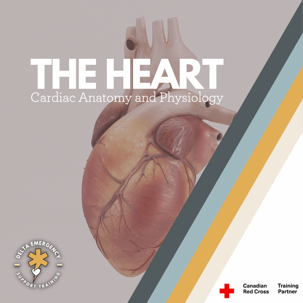
The human heart is a powerful, muscular organ central to the circulatory system, responsible for delivering oxygen and nutrients to tissues while removing waste. About the size of a clenched fist, it operates through a coordinated series of electrical and mechanical events that keep blood flowing efficiently throughout the body.

For Emergency Medical Responders (EMRs), a foundational understanding of cardiac anatomy and physiology is essential. The heart has four chambers (two atria and two ventricles), four valves that ensure one-way blood flow, and a conduction system that generates and coordinates each heartbeat. These components work together in two circulatory loops—pulmonary and systemic—to maintain oxygenation and perfusion.

Recognizing early signs of cardiac distress, using tools like ECGs, and performing interventions such as CPR or AED use all depend on a clear understanding of how the heart functions. From arrhythmias to cardiac arrest, EMRs are often the first line of defense in identifying and managing life-threatening cardiac conditions.

Read More

Essential Acronyms and Medical Terms Every EMS Professional Should Know

Essential Acronyms and Medical Terms Every EMS Professional Should Know

Mastering medical acronyms and terminology is essential for every Emergency Medical Services (EMS) professional. From understanding basic patient care concepts like ABCs (Airway, Breathing, Circulation) to advanced procedures and diagnoses, knowing the right terms can make all the difference in high-pressure situations. This guide highlights key acronyms and terms, alongside study aids and tips to help you quickly memorize and apply them in real-life emergency scenarios.

Read More

Meningitis: What Every First Responder Needs to Know

Meningitis: What Every First Responder Needs to Know

Meningitis is a serious infection that can escalate rapidly. As a medical first responder, understanding the types and symptoms of meningitis is essential for effective care. At Delta Emergency, we provide advanced first aid training across Canada, particularly in Edmonton and Calgary, to equip fire fighters and emergency responders with life-saving knowledge.

Read More

Understanding Medical Terminology: A Guide to Prefixes, Suffixes, and Roots for First Responders

Understanding Medical Terminology: A Guide to Prefixes, Suffixes, and Roots for First Responders

Mastering medical terminology is essential for first responders. Understanding common prefixes, suffixes, and roots will help you communicate more effectively, make informed decisions, and provide quicker, more effective care. In this guide, we break down essential medical terms that will boost your confidence on the job and improve patient outcomes.

Read More